On X-rays of children and adolescents, bones appear as if they have cracks—some with one, others with multiple. These “cracks” are actually cartilage that allows for the elongation and enlargement of bones during growth phases, known as epiphyseal plates or growth zones.
Juvenile Osteochondritis
Description
These cartilage zones, due to their mechanical characteristics, are softer than the surrounding bone and thus less resistant and more sensitive to mechanical stresses. On one hand, they provide additional elasticity to the skeleton, or parts of it, so single traumas are less likely to cause fractures, such as in falls or twists. This fact is not absolute; it depends on the intensity and direction of the trauma itself. However, on average, falls in children and adolescents less frequently result in bone fractures than in adults, precisely due to the added elasticity in bones during the growth phase and the presence of growth plates. This is a positive aspect since, by nature, children fall more often than adults, partly due to their adventurous spirit and partly because their neuromuscular system is still developing and testing its own limits. The downside is that growth plates are less resistant to small but repeated mechanical stresses, such as running and jumping, especially during the accelerated growth phase between ages 10 and 15. When physical activity, particularly sports, exceeds the growth plates’ capacity for shock absorption, adaptation, and recovery, an inflammation develops in them, which can cause pain for months and, in some cases, even years, occasionally lasting until the end of growth.
Inflammation is one of the most important bodily mechanisms of healing. Today, in popular literature, on the Internet, and on social media, inflammation is demonized, and advice is given on reducing or eliminating it. To this end, various supplements, special dietary regimens, and other methods are recommended, often claiming that inflammation is inherently bad and should be avoided or minimized. While there is some truth to this, trying to classify inflammation as simply “good” or “bad” and finding a pill or procedure for the latter prevents a full understanding of the complexity of the body’s adaptations to internal and environmental changes.
Put simply, inflammation is the process of healing injured or damaged tissue. For example, consider a simple cut on the skin. It heals in three phases.
The first is acute inflammation, characterized by the formation of a scab, redness around the wound, and pain. This acute phase involves numerous activities on both microscopic and macroscopic levels, aiming to prevent infection, close the wound with a scab as quickly as possible, and prevent the spread of the injury through mechanical, chemical (decomposition products of the injured tissue), and biological (bacteria and viruses) means. It lasts several days and gradually transitions into the second phase—subacute inflammation. In this phase, the primary healing of damaged tissue occurs. If the skin wound is small, skin cells will gradually multiply to close the injury. If it is large, healing will occur by forming connective tissue, which we call a scar. Initially, the scar is relatively large and red, as it is filled with many capillaries that supply the area with all necessary nutrients and oxygen for healing. Over a few weeks, subacute inflammation transitions into the third phase—remodeling. The connective fibers in the scar are initially laid in all directions and in an amount exceeding what is needed just to close the wound. This is the quickest way to restore tissue function. Once full function is achieved, the body gradually replaces the extensive scar with a smaller one, aligning connective fibers parallel to the direction of load or tension, and reduces the capillaries, which is seen as a change in the scar’s color from red to pale pink and, finally, to gray-white or the skin’s tone. Only then has the inflammatory process fully completed its task. All these phases, from the initial injury to the end of healing, are also called the inflammatory cascade.
From this explanation, it is clear that without the inflammatory process, we would not survive even the first year of life. It is the primary repair mechanism for tissue damage throughout our lives.
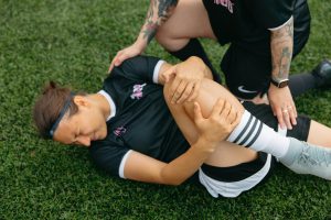
There is, however, a type of inflammation that is not beneficial, known as chronic inflammation. Simply put, it begins similarly to acute inflammation but stops halfway through. It is marked by hypervascularization (excess capillaries in the area), pain, and changes in the inflamed tissue, though not those that would lead to healing. It’s as if the inflammatory cascade has gotten stuck in a dead end, looping endlessly. These processes are undesirable anywhere in the body and can be triggered mechanically, as well as by various other factors, such as degenerative processes, autoimmune diseases, chemicals (like smoking), etc.
For this story about juvenile osteochondritis, the creation of chronic inflammation due to repeated physical stress is significant—in other words, a cumulative injury, or overuse syndrome. Bones are designed to withstand large amounts of small stresses over extended periods. The cartilage in growth plates is more sensitive to tension and pressure forces, and when these forces exceed its capacity for adaptation and recovery, inflammation develops. Due to the high metabolic activity of this tissue (after all, it is the “factory” of new bone), the increased metabolism from inflammation creates an environment where natural metabolic processes and the inflammatory cascade combine into a complex setting that does not lead to healing but exhibits aspects of chronic inflammation. This state is difficult to treat because rest reduces pain but generally does not resolve the issue completely, and the problem returns with increased physical activity.
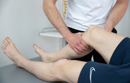
The most common sites for juvenile osteochondritis are the knee and heel, which is understandable as these parts endure the highest stresses during running and jumping. However, cases have also been observed in the hip, elbow, wrist, and shoulder, although much less frequently.
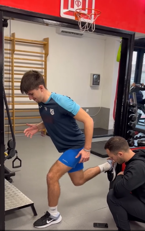
The treatment principle is similar for all and consists of three phases. The first phase is rest with physical therapy for 2–4 weeks. Then, if symptoms have decreased, we proceed with exercises within a pain-free range, gradually increasing the intensity to the young patient’s tolerance level. The goal of this phase is to “vaccinate” the inflamed tissue against mechanical stress, encouraging adaptation and reducing the inflammatory reaction during increased physical activity. This phase may last from several weeks to several months. After this phase, a gradual return to sports follows.
Unfortunately, this process does not guarantee complete resolution of pain. In the most fortunate third of patients, inflammation fully subsides, and it is possible to resume full sports activities. In the less fortunate third, a mild pain remains, allowing for limited sports activity, but not completely. In the least fortunate third, the pain returns despite the best-conducted therapy, and the only solution is to wait until growth is complete when the growth plates close and become bone. At that point, all pain disappears.

