
YOUR QUESTIONS AND OUR ANSWERS
YOUR QUESTIONS
AND OUR ANSWERS
- All
- Head and Neck
- Shoulder and Shoulder blade
- Spine
- Pelvis and Hips
- Knee
- Elbow and forearm
- Ankle and Foot
- Tension model
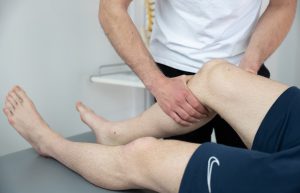
In a lengthy letter, a reader from a neighboring country describes his postoperative condition as good after knee arthroscopy and partial removal of the damaged meniscus, as well as detection of cartilage softening in certain parts of the joint, and asks:
What are the BEST glucosamine sulfate tablets (because looking online, there are various combinations…) or something else?
Is there a specific food or drink to speed up/improve the current situation? Are there any plants, is it true that calendula ointment and comfrey plant help? Injections?
Unfortunately, no active ingredient taken in tablet form or applied as a cream or compress has proven its effectiveness in terms of visible cartilage regeneration. Rare studies suggest possible reduction in pain and swelling with the intake of certain dietary supplements, but even then, not in all patients. Injections containing hyaluronic acid can be used, emphasizing that we also call them viscosupplementation.
Literally translated, this is “oil for lubrication,” which has no effect on cartilage regeneration. The approach we recommend for patients with early knee arthrosis is initially strengthening the muscles surrounding the knee. This is followed by long-term regular physical activity through a comprehensive program of strength, aerobic exercise, and proprioception with three main goals: -further strengthening of muscles -improvement of cartilage metabolism through aerobic exercise -stimulation of cartilage adaptation (as much as possible) through gradual increase in training load and proprioception.
We can say that this is not treatment in the strict sense of the word, but rather more of a prevention strategy aimed at slowing down degenerative processes and preserving full knee function. Our experience shows that this is achievable in most of our patients with early knee osteoarthritis. However, persistence is the most important factor, along with guidance from an experienced physiotherapist.
I am experiencing severe pain in my left shoulder. I have undergone 30 physical therapy sessions and received 7 shoulder block injections, but without improvement. MRI of the left shoulder shows: degeneration of the supraspinatus tendon, subscapularis tendon, and the intra-articular portion of the long head of the biceps brachii tendon. Osteoarthritic changes in the acromioclavicular joint with narrowing of the acromiohumeral space (possible impingement). Subacromial-subdeltoid bursitis. X-ray findings in the soft tissue projection cranial to the greater tubercle of the humerus reveal an oval mineral shadow 4 mm in diameter, characteristic of an intratendinous calcification. Enthesopathy of the greater tubercle of the humerus. Please explain these findings and advise on treatment.
Your findings point to moderate degenerative changes in the soft tissues of the shoulder, particularly in the rotator cuff tendons. This kind of MRI suggests the presence of extreme shoulder movements, with possible mild nighttime pain and discomfort during physical exertion. If the symptoms are significantly more severe than described, especially if they persist after repeated injections and long-term physical therapy, the underlying causes should be investigated beyond what is visible on the MRI and X-ray. This can be done through a detailed physical examination. There are many possible causes, with the following three being the most common in our clinic:
Frozen shoulder (adhesive capsulitis) – Inflammation of the joint capsule leading to the formation of scar tissue. Symptoms can last for years and are characterized by limited mobility and pronounced nighttime pain during the first few months. Typical physical therapy exercises and swimming often aggravate this condition.
Limited mobility of the cervical and thoracic spine, with or without direct nerve compression. Since the shoulder girdle functions as a unit that includes the spine, several joints around the shoulder, and muscles and tendons, reduced mobility in one part can provoke pain in another. Prolonged sitting and shoulder activity in that position can particularly exacerbate the discomfort.
Weakness in the muscles of the interscapular region, with or without scapular dyskinesia. This is typical for physically inactive people whose work involves sitting or standing for extended periods.
Each of these conditions is treated with specific physical therapy procedures.
One of our readers has been experiencing pain in both Achilles tendons for the past year and a half. He is typically very physically active and runs many kilometers per week.
X-ray imaging showed calcifications at the insertion points of the tendon into the heel bone (better referred to as osteophytes), and subsequently, shockwave therapy was performed (a total of 5 therapeutic procedures), along with stretching and attempts at rest, all resulting in transient improvements.
Like in most similar cases, he seeks advice on whether to repeat shockwave therapy, or if there is any “laser” or similar treatment that could relieve his pain and allow him to continue his activities in the same way that led to his pain.
Osteophytes on the heel bone are not uncommon. We regularly find them in the older population, and more frequently in extremely physically active patients. They themselves are not a problem (unless they are exceptionally large), but rather a sign of bone adaptation to significant and prolonged physical stress. In this sense, they can only be surgically removed. NO FORM OF SHOCKWAVE THERAPY CAN “BREAK” THEM, but only reduce local pain. If this was not successful, then the reservoir of simple therapies is empty, and a more comprehensive diagnostic evaluation and subsequent therapy are needed.
If it is clear that these osteophytes are a reaction to stress, then it is important to assess where this stress is predominantly occurring in order to reduce it. Typically, we find a lack of adequate warm-up and sufficiently prolonged stretching after running, as well as other training errors that contribute to pain and need to be corrected. This is followed by a physical examination, which will assess whether there are any changes on the foot and lower extremity that increase stress and can be corrected with manual therapy, orthotics, or changes in training.
Once all of this has been done, sufficient time is needed for such changes to bring satisfactory results. In this case, the guidance of an experienced sports physiotherapist is crucial.
In a lengthy letter, an eighteen-year-old volleyball player sought advice for her problem. Over the past year, she had occasionally experienced groin pain, which would spontaneously disappear or resolve completely with short physiotherapy, only to recur after some time. Initially diagnosed as a groin muscle strain, recent X-rays revealed hip dysplasia.
Her questions revolved around treatment methods and prevention of future pain, as well as the risks associated with playing volleyball with this diagnosis.
In sports physiotherapy, we are always searching for the underlying cause of symptoms that hinder an athlete’s normal training and competition. Muscle strains and partial ruptures are convincingly the most common reasons why young and older athletes seek the help of a sports physiotherapist. As these injuries are usually benign, relatively easy to diagnose, and treatable, we rarely conduct a full examination of surrounding joints, but we should. Processes within them can be direct causes of muscle pain that may appear as a strain but are not, or they can set the stage for muscle damage. We see dozens of cases like this girl’s every year.
Hip dysplasia is a term we use to describe a greater or lesser incongruity of joint bodies (the head and socket). In the hip area, it’s most often a shallow socket compared to the head of the femur. Such a condition may or may not cause pain.
In this particular case, the hip dysplasia isn’t severe enough to warrant a ban on sports activities. However, modifying training loads along with appropriate therapeutic exercises is a necessary change to make. Regular check-ups with an orthopedist and physiotherapist are necessary throughout the entire period of intensive sports activities. As always, a proper and specific diagnosis is half the battle on the road to injury recovery or pain management.
 A few years ago, I underwent therapy with you for a frozen right shoulder. After the therapy and exercises, I was feeling well. However, two months ago, the same shoulder started hurting again. Does this mean it is freezing again? I remember you mentioned that a shoulder freezes only once in a lifetime.
Frozen shoulder is the name for a condition that involves inflammation of the shoulder joint capsule, along with the deposition of connective (scar) tissue. The end result is pain and restricted movement. If left untreated, it usually lasts about a year and a half before it resolves on its own. Improper treatment, often involving aggressive attempts at mobilization, can extend the condition’s duration, as such interventions can prolong inflammation and slow the healing of connective tissue. With proper treatment, this unpleasant but benign condition can be shortened to an average of six months, according to statistical results from our clinic.
Generally, if the patient has no other conditions, the shoulder freezes (and unfreezes) only once in a lifetime. The only exception to this rule is in patients with diabetes, where the shoulder can freeze multiple times or may never fully unfreeze.
Another possibility in this case is that the shoulder has become painful for other reasons (issues with the cervical spine, bursitis, damage or inflammation of deep tendons, or other causes of pain and dysfunction). Therefore, it is advisable to perform a detailed examination to diagnose the current state and then treat it accordingly.
I would like to ask for your opinion.
I’m feeling tightness in the groin, specifically the adductor muscles.
Both sides tighten up, but more so on the right. It’s just one area that feels tight, and I feel tension there. I haven’t strained anything or had any injuries.
I stretch gently, but it doesn’t help at all. I’m quite active; I run, exercise, do leg and abdominal strength exercises, but this muscle can get very tense. I don’t know what else to do anymore.
There are four common causes of groin pain in physically active individuals – muscle injury (strain or partial rupture), tendon overuse syndrome of the groin muscles (tendinosis, enthesopathy, depending on whether the process is in the tendon itself or at its attachment to the bone), groin hernia (or colloquially, hernia), and hip impingement or arthritis.
From this brief description of symptoms, it’s only possible to conclude that it’s not a muscle injury because those typically have a clear and sudden onset, usually during a sudden movement or fall, which is not the case here.
It’s possible that it’s a condition related to the tendon or its attachment, but I would expect some reduction in physical activity and persistent stretching to be helpful.
Groin hernia (or hernia) may not be visible as a bulge. Even minor weakness in the abdominal wall can cause pain during more intense physical activity. It often comes with pain when coughing or sneezing.
Hip impingement is a condition where, due to specific anatomy, some movements are restricted, leading to the development of pain, primarily located in the groin region.
Which of these diagnoses is correct in the specific case can only be determined through a detailed examination, and the therapy radically differs for each of them. The shortest path to diagnosis starts with a simple X-ray of the pelvis and hips, followed by a physical examination, and possibly, if requested by an experienced orthopedic surgeon or physiotherapist, an ultrasound or MRI.
As always, I do not recommend self-diagnosis or treatment based on internet instructions or advice from well-meaning but unqualified acquaintances. It’s not uncommon for such actions to delay appropriate treatment, thereby prolonging it, sometimes significantly.
I’ve been actively involved in fitness for two years. About 6 months ago, I started experiencing pain and discomfort in my right shoulder. Sleeping on the right side became difficult, and some movements were restricted. An ultrasound of the right shoulder revealed “point-like calcifications at the insertion of the supraspinatus tendon into the greater tubercle with surrounding edema and a small amount of effusion in the subacromial-subdeltoid bursa, along with a discontinuous, hyperechoic tendon – consistent with a partial rupture of the mentioned tendon.
The biceps brachii tendon is of heterogeneous structure with minor linear effusions at the insertion site and more distally toward the myotendinous junction, indicating an incipient lesion of the mentioned tendon. I’m wondering what to do and which direction to take since I’m employed and actively involved in fitness?
In simplified terms, this is a cumulative damage to two tendons in the shoulder due to overuse. In such cases, it’s usual for us at Scipion to perform an ultrasound of the other shoulder to determine if there is a similar injury there, which may not be symptomatic. This is followed by a thorough examination of the neck, thoracic spine, and the entire shoulder girdle, as well as the scapulothoracic rhythm, to determine signs of restricted mobility, muscle imbalances in the form of local weakness or poor flexibility, and other disorders that may be causing pain. We also conduct a detailed “training process diagnostics” to get an insight into possible causes of damage within the training regimen. If the results of the examination and available diagnostic procedures are clear, we proceed to therapy. If there are uncertainties, additional diagnostics are indicated (most commonly MRI imaging). Pain therapy may involve various physiotherapy procedures, but it mostly relies on manual methods. Once the pain is alleviated, we provide precise instructions for modifying the training and teach the patient how to perform additional exercises to prevent recurrence of symptoms.
 For years, I’ve been struggling with neck pain that radiates down to my left shoulder, all the way along the spine between the shoulder blades. I’ve had an X-ray of my cervical spine taken, and I kindly ask you to explain the findings to me. Lately, I’ve been experiencing a kind of dizziness, a strange sensation in my head, and a tendency to sway to one side while walking.
Findings: Standard X-rays of the cervical spine reveal discreet dextroconvex scoliosis with straightening of the cervical lordosis. There are spondylo-deformative and spondylarrotic changes from C2 to C7. Osteochondrosis of the vertebral body endplates is observed. Narrowing of the intervertebral space at C4-C5 and more pronounced at C5-C6 is noted. There is discreet narrowing of the intervertebral space dorsally at C6-C7. Uncovertebral arthrosis from C3 to C7 is present.
Diagnosis: Bilateral cervical brachial syndrome.
The X-ray images indicate degenerative changes in the cervical spine, which are elaborately described. The findings do not directly indicate the cause of the problem, nor can X-rays do so (except in rare cases of fractures, tumors, and some others).
Symptoms of chronic vertebral pathologies, in this case neck pain, are associated with a variety of causes. A sedentary lifestyle, lack of physical activity, particularly adequate exercise, potential excess weight, and consequently poor circulation and lack of stimulation of bone and surrounding soft tissue metabolism, stimulate degenerative changes, which then occur more rapidly, comprehensively, and produce more symptoms. Simply put, everything in nature either grows or decays, including our tissues that we do not use.
The described discomfort can be alleviated with medication, sometimes responding well to conventional therapy (electricity, ultrasound, laser, magnet), but such improvements are transient. This is because degenerative processes have altered the shape and relationships between bones and soft tissues in the neck. Additional assistance in this case may include mobilization and manipulation techniques from the field of manual medicine. However, the most common permanent solution is regular exercise, which will maintain the mobility of the cervical and thoracic spine, stimulate circulation, and strengthen, and then maintain, the muscles to be strong and resilient.
Top of Form
a.
“I have read several of your articles related to myositis ossificans (a bone within the muscle), and I would need your opinion. I have been diagnosed with a calcification of 8mm in the belly of the triceps muscle caused by a hematoma that occurred on September 1, 2016.
I do not have any pain in the area where the calcification is, but it causes me significant discomfort in the form of a feeling of tightness in the elbow and noticeably weaker ability to use the triceps muscle, especially under pressure. According to the recommendation of an orthopedist, I underwent eight sessions of radial shockwave therapy, but I did not feel any positive effect after these sessions.
Currently, I am undergoing ultrasound therapy at the University Hospital Center Split. While reading one of your articles, I came across information that focused shockwave therapy has significant advantages over ultrasound therapy.”
A post-traumatic calcification in the muscle of the upper arm smaller than one centimeter should not cause any trouble. If there are symptoms present, it is much more likely that it is due to the connective tissue surrounding the calcification, which changes the direction of forces through the muscle, thus increasing tension in the elbow, rather than the calcification itself acting as a physical barrier. Shockwave therapy in calcifying myositis serves SOLELY for symptomatic treatment, meaning pain relief. It is NOT POSSIBLE to “break” such a calcification because it is not a buildup of calcium salts but rather a solid, live bone that has grown where it shouldn’t be. This means that we cannot expect its reabsorption after shockwave therapy. This reabsorption will occur spontaneously, but over a long period of time (several months, and sometimes years). In this light, the current ultrasound therapy also does not make much sense. If ultrasound therapy has not helped, it may be worth trying focused shockwave therapy, which can more precisely and deeply transmit energy with the aim of desensitizing local pain receptors.
However, more importantly than all of the above is to examine the shoulder and elbow and prescribe appropriate exercises to normalize muscle activation during movement and thus alleviate pain.
In a lengthy letter, a reader describes unbearable pain in the thoracic part of the spine that has been going on for two years and hasn’t subsided even with physiotherapy and chiropractic treatment.
She seeks advice.
In our practice, thoracic spine pains are not uncommon.
Most often, it is a so-called “facet syndrome” where there is reduced mobility of one of the small joints, causing pain, sometimes very acute. Conveniently, it either passes quickly without treatment or is easily treated with mobilization or manipulation techniques (chiropractic).
This is followed by pain due to soft tissue pathology, which encompasses a wide range of possible causes, all of which heal more slowly, requiring longer therapy and patient discipline, including regular exercise. Then there are the intervertebral disc herniations, which although much less common than those in the neck or lower back, can cause problems and require longer recovery. Sometimes, despite the efforts of doctors, therapists, and patients, the pain does not decrease or returns repeatedly. Interestingly, this phenomenon, so specific to the thoracic spine, has been known for a long time and was detailed by the pioneer of manual therapy in Europe, Dr. Cyriax, back in the 1960s.
In such cases, a classical clinical examination is not sufficient. Additional diagnostics are necessary, primarily referring to an MRI. It should show the condition of the discs, small joints, and the muscles surrounding the spine. If it does not indicate a probable cause of the pain, additional specific examination of the mobility of the scapula and cervical spine is necessary to see if there are any “blockages” in a dynamic context that irritate the sensitive soft tissues. Sometimes a neurological examination and EMG are required, and depending on the case, the internal or pulmonary status should be determined by qualified specialists, as some lung or internal organ diseases can show symptoms as middle back pain.
Only after the diagnosis is established can an individual therapeutic approach be created with promising possibilities for success.
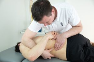
 Recently, a football player sought help a few days after sustaining a thigh injury during a match. Two days after the injury, the front muscle of the thigh was painful and slightly swollen when a rough massage was performed at the club, colloquially called “breaking up the hematoma.” The day after the massage, the thigh swelling increased, leading to the hematoma being drained via puncture.
During an examination in our clinic, an ultrasound revealed a significant muscle rupture accompanied by swelling. Such injuries typically require several weeks of rest from sports activities, along with appropriate physiotherapy.
But what about the “breaking up the hematoma” technique? This concept is very old and has been part of sports physiotherapy since the field’s inception. The logic behind this therapy is that the local hematoma (blood trapped in the tissue as a result of a ruptured or contused blood vessel) should be encouraged to spread over a larger area to facilitate easier resorption. This rationale is rooted in physiology, and when done correctly, it can significantly reduce overall recovery time and speed up the return to sports. The issue lies in the timing and intensity of such a massage.
To ensure that the injured blood vessel has healed, time is needed—at least 48, preferably 72 hours after the injury. If the blood vessel hasn’t healed, any massage, especially a rough one, can trigger further bleeding, increase the hematoma, and worsen the injury, which is likely what happened in this case.
A particularly rough massage adds further trauma to all the tissues under pressure, causing reactive swelling, even if not visible to the naked eye. Such traumatized tissues around the hematoma are less able to resorb it, simply because they are also injured and focused on their own healing.
The stimulation of post-traumatic hematoma delocalization (its spreading over a larger area) through massage and other manual techniques should be done with light to moderate pressure over a prolonged period. When performed this way, the therapy lacks a destructive component and directly reduces recovery time.
 “I am a 50-year-old healthcare worker, and I have a long-standing condition in my right knee in terms of femoropatellar joint arthritis and grade 2 arthritis of the internal joint bodies. In a recent knee MRI, there is a finding of anterior cruciate ligament rupture which I believe is a result of an injury from 30 years ago. I have been involved in recreational sports, and for the past 8 years, I have been playing tennis. I have read your advice on strengthening the thigh muscles. I do not have the support of my doctor to continue playing tennis recreationally. My question is, can I strengthen the muscles and stabilize the knee to the extent that I can continue to play lightly, or is that impossible.”
Generally, with existing knee arthritis, sudden accelerations, decelerations, and changes in direction, especially in situations of anterior instability, pose risks for further cartilage damage and worsening of the condition. That is why in the medical profession, we do not like it when patients with arthritis play tennis. However, in my clinic, there are many patients like you. For them, a certain sport that they engage in with particular passion represents an important part of their overall quality of life, and they feel bad without it. Some of them are willing to accept the risk of faster progression of their degenerative diseases in exchange for the opportunity to continue engaging in activities that make them happy. If you are one of those, here are some tips to reduce overall stress on the knee and its cartilage:
Find an experienced sports physiotherapist in your area who will guide you, determine the level of load in training, give you an exercise program, persuade you to take occasional breaks, and assess the need for physiotherapy. Prefer doubles over singles, as the loads on the knee are lower. Regular low-intensity aerobic training (static bike, elliptical trainer), and regular stretching should become your best friends. Several days of complete rest each month must become routine.
Recently, a young Hungarian javelin thrower sought help.
Her issues with her right shoulder began a year ago, and in the meantime, she underwent appropriate therapy at another medical facility, which reduced the symptoms. However, it did not completely eliminate them.
Despite regularly doing prescribed exercises, she still cannot throw without pain, and therefore cannot achieve the results she aspires to.
The examination showed no clear inflammatory process or tissue damage, and the pain was related to the less mobile right scapula. The way her scapula moves during arm movements indicated that, at this moment, the so-called stabilization exercises, which initially helped her reduce pain, caused a new problem that now troubles her. By changing the exercises and providing additional education on how and when to perform them, the issues disappeared, and she is now training at full intensity without pain.
Many similar stories could be told, ranging from benign to more serious problems caused by therapeutic procedures that were either not discontinued in time or applied in entirely inappropriate situations. Lately, we most often see them as a result of self-attempts at treatment, following advice obtained from the internet or from individuals who have had “the same problems” and feel confident enough to give advice based on personal experience.
Every clinician learns early on the simple truth that therapy or medication is only effective if applied in precisely defined circumstances and only for as long as necessary. Even then, success is not always guaranteed. Differences between patients, which may not always be clearly visible or seem important, affect the effectiveness of therapy. The protocols we follow in determining therapy are merely recommended directions that must be adjusted to the specific case. Monitoring the therapeutic effect is of paramount importance because the body’s reaction indicates whether the direction of treatment is appropriate or needs to be corrected. Therefore, expert guidance in any treatment is essential.

“I awkwardly stepped during a futsal game and immediately felt a slight popping sensation in my left knee. The pain was instantly intense, and within a few minutes, I managed to stand up but limping. The next day, I could hardly put weight on my left leg, and swelling appeared around and above the knee. The swelling decreased after 5 days. After 15 days, there is swelling again, although I have no pain when moving, although it’s not like my right leg. Can the fluid, if present in the knee, just withdraw, or do I need to have it drained? I’ve tried various home remedies, but ice helps the most, and wrapping it with foil overnight.”
Swelling of any joint can be twofold. One is sudden swelling after a stronger sprain or impact. It mainly consists of blood that fills the joint space, coming there due to blood vessel injury. After 2 days from the injury, it is highly recommended to puncture such a joint (using a needle and syringe to remove as much blood as possible from the knee), because blood from the joint is resorbed very slowly, and additionally, it can adversely affect the cartilage. The other type of swelling occurs more hours to two days after minor sprains, or without a clear cause. If we puncture such a joint, we will get a yellowish fluid without blood. It is pointless to puncture or re-puncture such swelling because it will reoccur very quickly, within a few hours. It usually represents a defensive reaction. Namely, if there is damage to the cartilage (or probably the meniscus in this case), the body will try to reduce friction between the damaged parts by placing fluid between them through swelling. In such cases, a good diagnosis is half the battle, and the patient’s understanding of the causes of their problems is the other half, because all subsequent physiotherapy and exercise procedures require some time and patience. Thus, in the case of cartilage damage, which is not large enough for surgical intervention, it is possible to “desensitize” the joint through an individual kinesiotherapy program and prepare it for sports activities within a reasonable period.
Can you please give me your opinion? I injured my arm two months ago at a meat processing plant. Initially, my arm started tingling, initially less, for about half an hour to an hour a day, then the pain started and the tingling increased. I went to the doctor, and he told me that a nerve or tendon was inflamed.
I had an EMG done, which showed that the median nerve in my hand was moderately damaged. The doctor prescribed 12 injections: 10 injections of vitamin B12 and two injections that I received every seven days. Also, when I twist my hand, the middle finger hurts a little as if it’s not mine, and the bottom fourth finger feels a bit tingly when touched. When I make a fist and try to pull it upward, it tightens and hurts me, and it feels like something is clicking in my shoulder.
Can you give me advice on what to do and how to proceed? I also need to go abroad for work at the end of March. I need to recover by then. Can you give me advice on what to do? Thank you in advance.
From the description of the symptoms, it’s not clear whether it’s carpal tunnel syndrome or cervical brachial syndrome (pressure on the nerve as it exits the neck). Regardless, medication therapy is necessary but not sufficient. If it’s carpal tunnel syndrome, in addition to laser and ultrasound therapy, it’s advisable to undergo manual therapy focusing on mobilizing the joints of the hand and forearm, with special emphasis on mobilizing soft tissues and stretching exercises.
If cervical brachial syndrome is at play, in addition to neck and shoulder massages, traction of the neck is highly recommended, as well as mobilization techniques for the cervical and thoracic spine.
I emphasize that for complete recovery and the speed at which it will occur, the patient’s patience is crucial because nerve recovery is slow and can take months. Additionally, after completing therapy, it’s extremely important to continue exercising regularly to prevent the recurrence of symptoms.
I would like you to explain this finding to me and how it is treated.
Conclusion: Lordosis. Left scoliosis at the level of the C6-C7 segment. Status after ligament injury at the C6-C7 segment with dorso-medial protrusion at the C5-C6 and bilateral paramedial protrusion of the intervertebral disc, causing compression of the C6 nerve root bilaterally.
One of the world’s most renowned shoulder surgeons, a Frenchman by origin, consistently emphasizes that he operates on patients, not images, in response to such questions. By this, he means that not only surgery but orthopedic medicine in its entirety, including physiotherapy, can and should treat the patient’s symptoms, rather than the changes seen in imaging diagnostics, in this case, an MRI.
A comparison that resonates with my patients is a common blister on the heel. If you get one, the first thing you’ll wonder is what caused it. Usually, it’s the shoe. Whether it’s too tight, too loose, tied too tightly, or inadequately fitting in some other way, it will cause a blister that hurts. Similarly, in this case, images of the blister itself can describe it in detail, but they cannot indicate what led to it, let alone what to do to alleviate or remove the condition. Because if you go to a podiatrist, they remove the blister, but if you continue to walk in the shoe that caused it, nothing is accomplished.
In this specific case, it involves a complex change in the cervical spine, including disc deformation, the spine itself, and associated ligaments, which may be partially caused by trauma, exacerbated by degenerative changes. The images we see cannot be changed by therapy or surgery to resemble the state before the injury or ten or more years ago. What is possible, however, is to examine the patient, assess the condition of other muscles, joints, and other parts of the musculoskeletal system, and through stimulation of compensatory mechanisms abundant in the body, reduce or eliminate pain, and establish normal, or at least satisfactory function of the cervical spine, and subsequently, the body as a whole.
A few weeks ago, a physically active 22-year-old young man sought help at our clinic, who has been experiencing issues with his right shoulder for the past two years in the form of clicking during workouts at the gym.
Several examinations were done by various specialists, and an ultrasound indicated the presence of a small calcification in one of the deep tendons of the shoulder. He underwent physiotherapy and shockwave therapy to reduce symptoms and remove the calcification.
Despite all this, and as he did not improve, a magnetic resonance imaging (MRI) scan was performed, which, apart from a minor calcification, showed no other pathology.
During the examination, it became clear that the clicking occurs when initiating scapular movement while raising the arm. This condition is sometimes referred to as subscapular bursitis by some authors, but it has other names as well. Essentially, the scapula slides over the soft tissues covering the chest during movement. If these tissues become irregular, usually as a result of microtraumas typical of weight training, a condition characterized by crepitus, or clicks, can develop, as in this particular case. Some manual therapy and specific exercises significantly reduced symptoms in this young patient, and if he follows training advice, it is possible that they may disappear completely.
In an age where imaging diagnostics are the gold standard in diagnosing musculoskeletal conditions (ultrasound, MRI, CT, X-ray), there are still conditions that cannot be identified without a detailed examination and will not show up on the mentioned tests.
Furthermore, clicking in the shoulder girdle during movement can also be a sign of shoulder instability, degenerative joint changes, partial tendon ruptures, cervical spine changes, as well as some rare diseases. Each of these problems requires specific treatment and possibly additional tests to confirm the diagnosis and assess the level of damage. However, it all begins with a detailed physical examination.
I have chronic pain in my right Achilles tendon. It has been ongoing for two years. I’ve tried medications, physiotherapy, stretching, warming and anti-inflammatory creams. Nothing helps. I run several kilometers every day and occasionally play football. I’m afraid that my tendon might rupture, just like it happened to my friend a few years ago. What can I do to prevent that?
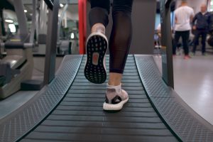
This is a very common question from my patients with Achilles tendon inflammation who are physically active. The simple answer is that the chance of an inflamed Achilles tendon rupturing is actually very small. The very fact that pain is present reduces the load during walking and sports activities in such a way that the body spontaneously shifts a greater overall load onto the uninjured limb. It is possible, for example, to walk in a way that distributes 60% of the load onto the painless leg compared to 40% on the injured one, without it manifesting as a limp or a visible change in walking or running rhythm. In this way, pain becomes a protective mechanism.
The vast majority of Achilles tendon ruptures occur without prior pain. This happens because a part of the chronic degenerative changes in the tendon can occur without pain. When there is no pain, there are no protective mechanisms, and without them, the risk of additional damage is very high.
However, despite your poor experience with physiotherapy so far, I recommend that you find an experienced physiotherapist in your area who will thoroughly examine not only your tendon but your entire lower extremity, hypothesize about the cause of the chronic inflammation, and work with you on interventions that should lead to the final healing of the chronic damage, and consequently, the cessation of pain.
Continuing sports activities with pain puts you at risk of developing new overuse syndromes in other parts of your lower extremities or spine.
I currently have a frozen shoulder from an accident that happened 1 month ago, and I wore a sling for 1 month. So, my arm didn’t move at all.
Which creams should I use to warm up my shoulder because it feels good when I exercise, but when it cools down, I endure the pain??
Stiffness in the shoulder after trauma and immobilization is not uncommon. As the most mobile joint in the body, any restriction of movement in it is clearly noticeable and interferes with daily activities. Particularly pronounced stiffness, accompanied by pain, especially at rest, during nighttime rest, or after exercising, is also known as frozen shoulder.
Like the “regular” frozen shoulder, the post-traumatic version is characterized by inflammation of the shoulder joint capsule, which causes pain, as well as the formation of scar tissue, which restricts movement. The cause in this case is trauma (impact, sprain, bone fracture, surgery, and even, although very rarely, injection).
For a long time, the standard approach in the field was that mobilization, especially with aids like a stick, together with rather painful stretching by a physiotherapist, was the main therapy. Relatively recently, a large Dutch study, which was later replicated, showed that such treatment prolongs the duration of the condition by 60-100%. From my practice, I see that this knowledge has not yet found its way into the standards of frozen shoulder treatment. In that sense, the dominant advice is still for the patient to exercise as aggressively as they can tolerate independently, while managing the resulting pain with medication, creams, or ice. Such approaches can be considered outdated, and although they seem logical, they only bring harm.
The modern approach is based on pain control (using various procedures from the field of conservative orthopedics and physiotherapy), manipulation of soft tissues to reduce global tension around the shoulder, and exercising exclusively below the pain threshold. In this process, the guidance and control of a physiotherapist are important, as well as the patient’s understanding of the nature of the problem and the time needed for physiological processes to lead to complete resolution of stiffness.
Top of Form

Four years ago, I injured my knee (anterior cruciate ligament rupture), and about a month ago, I injured it again, leading to a rupture of the posterior horn of the medial meniscus.
I’m wondering if the only solution for me is knee surgery since it’s now more unstable than before, or is it possible to improve the condition with some physical therapy?
The anterior cruciate ligament is an extremely important part of the knee stabilization system. Without it, there is micro-instability during movement, which over time can cause cartilage degradation and the onset of osteoarthritis. Intensive physical activities or sudden movements under load cause clear episodes of instability, which have the potential to damage the meniscus.
In this specific case, the expression “my knee is more unstable than before” points to the real problem. Meniscus damage is just its symptom. In that sense, it is possible to attempt physical therapy to alleviate the pain. Depending on a whole range of factors, it can be very successful. In case of its failure, it is also possible to undergo arthroscopy, during which the injured part of the meniscus would be removed, thus reducing the pain. However, the underlying problem of knee instability would remain untouched, as well as the risk of future similar injuries.
There are two possible approaches to addressing the symptoms of an unstable knee. The first is conservative. It involves a list of sports and physical activities to avoid, combined with regular exercise aimed at at least partially compensating for the missing ligament stabilization with muscle strength. The good side of this approach is avoiding surgery and long rehabilitation. The downside is living with significant limitations.
The second is surgical reconstruction of the anterior cruciate ligament. The surgery itself is now a routine procedure, but recovery from it takes up to a year. The good side of surgery is a life without restrictions in the end. The downside is the long rehabilitation and possible complications (now very rare), as with any surgical approach.
“After having an MRI of my right knee, the conclusion is a degenerative change in the meniscus with horizontal and vertical rupture of the posterior horn of the medial meniscus.
I work as a merchant and have terrible knee pain. After standing for an hour or two, I feel like my knee has stiffened.
Please give me your opinion on whether surgery is necessary or not. I am 45 years old.”
The menisci in the knee serve two roles. One is to improve knee stability by forming a deep plate that prevents slippage of the joint. In this sense, the joint socket becomes deeper, and the slipping of the head is limited.
The other role is cushioning. When the outer (lateral) meniscus is removed, studies have shown that the stress on the articular cartilage increases 2-3 times.
Surgical procedures on the meniscus are performed arthroscopically and can involve simply removing the damaged part or the entire meniscus. Alternatively, it is possible to attempt to stitch the meniscus, but this solution is suitable for very young people and specific injuries, and even then, some luck is needed for healing to occur.
In this particular case, it is clear that stitching will not be beneficial. If the menisci are indeed causing problems, then the only solution is to remove the damaged parts. The question here is whether these injuries are causing pain or if initial degenerative changes in the joint and cartilage are already at play, causing the damaged menisci to stand out the most on the MRI. To determine the answer to this question, a thorough examination needs to be conducted, and at least one attempt at physiotherapy aimed at improving local circulation and muscle strength should be made. If such a procedure sufficiently reduces the symptoms, the need for surgery ceases. Namely, except in very rare cases, surgery for meniscus cannot be delayed. This way, we avoid unpleasant situations where the condition after meniscectomy in terms of pain and other discomforts is the same as before surgery. Because it is entirely possible that the meniscus damage does not cause pain.
While browsing websites in search of information about Achilles tendon injuries, I came across your article…
Namely, I have exactly the symptoms you described (pain at the attachment of the Achilles to the heel), clearly without any “cracking” or thickening of the tendon. I am involved in recreational running (more trails than roads), and the pain began to occur somewhere in May. By the end of June, I stopped any training and went for physical therapy + rest throughout the summer (only light swimming and bathing). After all that, I see no improvement. For the past month, I’ve been running again twice a week (not too much, about 5-6km per session), and the pain is always present, but no worse than during the period without training. The long-term plan is to start with more intense training and preparations for races of ultra distances over 50km.
Your article somehow gives me hope that I have discovered the essence of the problem, I just don’t know how to solve it.
At the last professional seminar on applied or “functional” anatomy, one of the leading European authorities in the field, when asked how far the Achilles tendon extends, coolly responded: “If you are referring to the right Achilles tendon, then it begins in the vicinity of the left shoulder.”
In this column, we don’t have enough time for a detailed explanation of this statement. Suffice it to say that the forces generated during running partially extend all the way from the back muscles, through the hips into the lower extremity via intertwined connective structures. This means that, in principle, a problem in the back could be the primary cause of Achilles tendon pain, and vice versa. However, it is important to understand the significant difference between “could be” and “actually is”. The former is a question before examination, and the latter is a statement after examination and any necessary diagnostics.
All my texts are not a substitute for appropriate examination and therapy but rather inspiration and encouragement to seek them out.
I am 60 years old and have a kyphotic posture, which of course did not develop overnight. I bought a posture corrector belt that supposedly activates muscles and corrects posture. I’ve been wearing it for the past two days, but something doesn’t feel right to me.
Therefore, I kindly ask for your opinion on this matter. I don’t feel very good in it; it feels tight from all sides, my back muscles hurt, and I feel like it pushes my head even further forward.
I’m afraid it will do more harm than good. Maybe I’m wrong, maybe I need to get used to the belt. Your opinion is very important to me, so if you can spare a few minutes for a brief answer, I would be truly grateful.
There are no braces or belts for correct posture (it’s all marketing). Corrective braces used for scoliosis are effective, but only during the rapid growth phase, i.e., in adolescence. Everything after that is just torture devices that, at best, will bring no changes, and in all others, at least transient pain. Prolonged wear of such products directly weakens the muscles of the neck and thoracic spine and restricts the mobility of the small joints in them, which means they cause new disturbances in the long run. This happens in the same way that muscles in a leg immobilized by a cast weaken due to, for example, a fracture. Some of these braces, worn briefly during acute pain, can be helpful, but they are not corrective and cannot change the condition of your spine.
Therefore, changes in the shape of the spine cannot be corrected after the end of growth (except surgically). However, pain and discomfort can be alleviated through physical therapy methods, manual therapy, and especially through proper and regular exercise. Therefore, I recommend that you find an experienced physiotherapist in your area who will examine you and advise you on how to best help yourself.
On an MRI, they found a rupture of the anterior cruciate ligament. The orthopedist I consulted with said that I should have surgery and have a new ligament (made from tendons) put in. My knee only hurts when I squat or kneel for a long time. I read in one of your articles that surgery is not always necessary when this ligament tears. What do you advise me?
Patients with a rupture of the anterior cruciate ligament are divided into two large age groups. The first group consists of young people up to 30 years old, for whom we recommend surgery regardless of their current condition. Prolonged knee instability resulting from the lack of this important ligament almost inevitably leads to degenerative changes, and if they occur very early in life, their consequences can be such that treatment is difficult, requiring multiple surgical procedures and prolonged rehabilitation. The second group consists of patients aged 40 and older, for whom the most important question is not whether they have the ligament, but whether they have episodes of instability in their daily or sports activities. This condition is called manifest instability, and those who have it know that it significantly reduces their quality of life, so surgery is advisable for them. If such manifest instability is not present, it is possible to live entirely normally without surgical intervention, with appropriate exercise. Patients between 30 and 40 years of age are subject to an individual approach, and the decision is made on a case-by-case basis, with an important consideration being the type of sport the patient engages in. If it involves sudden changes of direction at full speed, or if it is a contact sport (soccer, skiing, basketball, tennis…), surgical stabilization would be advisable.
I myself lost my anterior cruciate ligament during an indoor soccer game when I was 37 years old. Since I can do without soccer, and satisfactory physical activity includes running and the gym, I was not operated on, I exercise regularly, and to this day I have no significant problems with that knee. If soccer were my passion, without which I couldn’t live, the outcome would be different.
An X-ray of the right shoulder taken on August 20th, provided on a CD (I do not have the report as the doctor is on vacation), shows a calcification approximately 2-3 cm in size as determined by a general practitioner. I would like your opinion.
Treatment depends on the following factors: size, shape, location, phase, and symptoms.
Size:
- Calcifications up to 1 cm are considered small,
- 1-2 cm are considered large,
- Over 2 cm are considered very large.
- The larger the calcification, the less likely conservative treatment will be effective.
Shape:
- Is the calcification thin and elongated, or round?
- In the first case, if it is within the rotator cuff tendon itself, it rarely causes significant problems.
Location:
- Front (in the subscapularis tendon): Usually causes intense but relatively short-lived symptoms that respond well to physiotherapy and exercises.
- Middle (in the supraspinatus tendon, most common): Smaller calcifications are successfully treated conservatively. If located in the bursa, they respond poorly to classic therapy but better to shockwave therapy.
- Back (in the infraspinatus tendon): Rare and cause fewer issues or are found incidentally during ultrasound examinations.
Phase:
- Every calcification has three phases: formation, maintenance, and resorption.
- In the first phase, pain is well managed with blocks.
- During the maintenance phase, shockwave therapy may help.
- In the resorption phase, classic physiotherapy is beneficial.
Symptoms:
- We often see very large calcifications causing minor issues and relatively small ones causing significant pain.
All these factors guide treatment towards:
- Medication therapy: For acute pain that is continuous and occurs at night.
- Classic physiotherapy, manual therapy, exercises: For subacute pain, not occurring at night, mainly during specific movements, in the second and third phases of the calcification’s life, and not highly irritable.
- Shockwave therapy: For calcifications that periodically cause flare-ups, with pain never fully subsiding, and when other treatments have failed.
- Barbotage (“washing out the calcification”) and surgical therapy: When the calcification is too large or after unsuccessful conservative treatment.
All these rules have many exceptions.

“Hello, I am a professional football player. I play in Asia. For the past four months, I have been dealing with a groin injury that radiates to the lower abdomen. Overall, I don’t know what caused the pain or what could be the underlying cause. I only know that it’s a significant problem for me to enter into duels at 100%, as well as sprinting. I have much less aggressiveness, and I believe that the injury is the reason for it all. Here, they are not in favor of me getting an ultrasound, as they believe that nothing will be seen. I also do exercises for groin warm-up before every training session. I take painkillers before every match, and then I generally feel better for the next two days, and then it starts all over again. I’m interested in what the smartest thing to do is. I’m from Serbia, and I’m considering going to have groin surgery if necessary. Because here, I would never allow them to do it. I would be grateful for any advice.”
The exact cause of chronic groin pain is not always possible to determine with 100% certainty. Therefore, collaboration between an orthopedist, a physiotherapist, and an abdominal surgeon is crucial. In football players, classical inguinal hernia is very rarely seen, meaning there is extreme laxity of the abdominal wall and protrusion of abdominal wall contents. Pain can be caused by weakness of the same wall, which then causes pain during intense exertion. In such cases (most common in football), weakness cannot be proven by physical examination, ultrasound, or magnetic resonance imaging. On the contrary, such a diagnosis is made after examination and diagnosis by an orthopedist and after appropriate physiotherapy. If there is no improvement after 3 months of persistent physiotherapy and focused exercise, and if hip MRI does not indicate other biomechanical disorders or inflammatory processes in the tendons, it is time to visit an abdominal surgeon. If other conditions are met, he will proceed with preoperative preparation and assess which surgical approach is best in the specific case, with the aim of stabilizing and reinforcing the abdominal wall. The two most common approaches are classical tissue suturing and mesh implantation, each with its advantages and disadvantages.
“I am involved in football, and I have been dealing with shin splints problem for 3 years now. I feel pain in both legs, more intensely in the left one. I don’t feel any pain while walking, but as soon as I start with faster movements than walking, the problem arises. I have been struggling with it for 3 years, and during that period, I have tried all sorts of therapies. Massages by physiotherapists, shockwave therapies, chiropractic treatments, energy therapies, body relaxation therapies, ice, mud, hot-cold water, video analysis of running, movalis tablets, etc., all unsuccessfully.
When there is a break between half-seasons, I take a rest, and after the preparations for the season start again, I initially have no pain, and then after some time, they reappear. I don’t know what else to try and do?”
The so-called shin splints syndrome is an overuse injury and represents the accumulation of micro-injuries to the tissue, which the body is unable to repair within the available recovery time. In this specific case, the localization of pain and inflammation, as a result of overuse, is on the inner side of the shin. The tibialis posterior muscle, which is the center of this problem, is responsible for balance and maintaining joint stability during running. Two facts are evident from the description of symptoms. First, local therapy does not yield results. The second is that the symptoms completely disappear after a longer rest. This latter fact is particularly important because it indicates that permanent changes in the tissues have not occurred, despite the duration of the symptoms. Cumulative stress can be reduced in this case in two ways. The first involves an examination to assess the mobility of all joints in the lower extremities (hips, knees, ankles) because stiffness in one or more of them directly and significantly increases stress on the tibialis, and then focused exercises aimed at eliminating that stiffness (from our experience, the hip and ankle are most often problematic). The second is a change in running technique in collaboration with an athletic trainer specialized in sprinting. Although it sounds simple, changing the way you run in sprint or change direction requires time and focus, as well as expert guidance.

My latest X-ray report of the lumbosacral spine states the following:
“Sinistroconvex scoliosis of partially biconcave formed vertebral bodies of the lumbar spine segment. The L2 vertebral body is lower in height. According to Genant classification, it is grade 1. Intervertebral osteochondrosis. Reduced intervertebral space at L5-S1. Reduced joint spaces of the small joints from the L3 level downward. Sclerosis of the spinous processes. Reduced joint space of both SI joints with enthesopathy of the iliolumbar ligament.”
I noticed that your clinic has introduced a new device for piezoelectric focused shockwave therapy, which is extremely precise (focus 4×6 mm) and allows adaptation of penetration depth, so I assume that you may be able to offer appropriate therapy regarding my findings.
The X-ray findings of your spine indicate degenerative changes, also known as spondylosis. These changes result from a combination of three factors in varying proportions — genetic predisposition, age, and lifestyle. Since the first two components cannot be controlled or changed, the third remains available for intervention.
These interventions consist of two groups of procedures — learning proper sitting posture and lifting techniques, as well as regular exercise aimed at reducing stress on the damaged parts of the system during movement, thereby reducing or eliminating pain. All of this assumes that your primary issue is lower back pain, with or without radiation into the lower extremities. Additionally, during the recovery process, other methods from the arsenal of physiotherapy may also be used as supportive treatment.
Shockwave therapy is a powerful technology for the treatment of chronic tendon inflammation, tendon insertions, and other connective tissue injuries, along with several additional indications outside those described above. We also use it as a purely analgesic technique.
Around 20 years ago, studies were conducted on chronic lower back pain, and their results were not particularly good. Around the same time, we also attempted to apply shockwave therapy to chronic lower back pain in several of our patients, with ambivalent results. The treated area is rich in nerves, which do not respond well to this treatment method. Although we did not observe significant worsening in our clinical practice, the short-term improvements achieved did not justify the use of this method. As described earlier, there are better and more effective treatment protocols.
Additionally, the sound pulses of shockwave therapy cannot penetrate deeply enough to reach the intervertebral disc itself or its surrounding structures, which are the locations of most pathognomonic changes in spondylosis. On the other hand, the small joints of the spine are within reach of this therapy and often respond well to it. Therefore, in a smaller proportion of lower back pain cases caused partly or predominantly by pathology of the small spinal joints, this method may be considered as part of treatment, but not as the sole form of therapy.
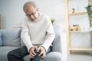
I am 45 years old man, and for the past two years, I have been experiencing occasional pain in both knees.
In the last two months, my right knee has been hurting more, so I went for an examination, and the orthopedist told me that I have moderate osteoarthritis. I searched the internet and saw that stem cells can be injected into the knee, which can repair cartilage.
Do you have any experience with patients who have received stem cells?
Osteoarthritis is a complex degenerative disease that affects not only the cartilage but also the bone beneath it. For easier understanding, we can compare cartilage to grass on a lawn, and bone to the soil underneath it. Just as the soil provides the grass with necessary moisture and nutrients, the bone through its blood vessels provides most of the necessary nutrients and oxygen to the cartilage. If it is not permeable enough, the cartilage will suffer. And that is precisely what happens in osteoarthritis. The first signs of this disease are visible on regular X-rays in the form of a bright line on the bone beneath the cartilage. This condition is called subchondral sclerosis and indicates a process of literal thickening and hardening of the bone on which the cartilage sits. Such hard bone increasingly fails to pass nutrients, resulting in faster “wear and tear” and slower recovery of the cartilage. The comparison with the lawn here is excellent. If we have concrete under the grass, you can pour all the new grass seeds and fertilizers you want, but it cannot be the lawn we knew.
In this sense, research on the effects of stem cells on osteoarthritis has mainly been conducted on a small number of subjects, and we do not have a definitive prescription for at what stage of osteoarthritis and whether this treatment method is effective at all. Even the authors of studies claiming significant benefits admit that they last for a maximum of 2 years per therapy. If you decide to undergo such treatment, do not forget that the only proven procedure for osteoarthritis, confirmed in tens of thousands of patients, which reduces pain, reduces doctor visits, and postpones surgeries, is regular and tailored physical activity, i.e., recreational sports.
Dear Sir/Madam,
do you provide therapy with focused electrohydraulic shockwaves for stem cell regeneration aimed at restoring cartilage in a hip affected by arthrosis, accompanied by pain and reduced mobility?
If so, which device do you use, what is the treatment schedule, and what is the price?
The device we use produces shockwaves through a piezoelectric system and differs from the electrohydraulic system primarily because it has a smaller focus. In this way, most of the shockwave energy can be delivered directly to the site of injury while reducing irritation of the surrounding tissues. There are also other differences, but they do not affect the physiological tissue response or treatment efficiency and are mostly technical in nature.
If you allow, I will simplify and reformulate your question as follows: Does shockwave therapy stimulate cartilage regeneration in joints?
The answer is simple — no. This therapy cannot do that, just as no currently available treatment method can stimulate the regeneration of joint cartilage, regardless of marketing claims, influencers, social media advice, or AI-generated instructions.
Shockwave therapy may help reduce pain in some patients with arthrosis through two mechanisms — hyperstimulation analgesia (pain reduction immediately after therapy) and stimulation of the inflammatory cascade in surrounding soft tissues, which can be responsible for a large part of the pain associated with arthrosis. Such a procedure also carries a risk (although not a major one) of temporarily increased pain, and the patient should be informed about and prepared for this possibility.
We use shockwave therapy for arthrosis only as part of a broader treatment plan, never as a standalone method, because our clinical practice has shown that on its own it is unable to produce the level and duration of pain reduction and mobility improvement that we aim to achieve in our patients.
I engage in recreational running and go to the gym.
For a year now, I’ve been having issues with the attachment of the Achilles tendon to the heel. The pain is present all day, stronger in the morning and after training.
My heel appears swollen. I’ve been to physiotherapy, wear insoles, and a month ago I even had shockwave therapy.
Nothing helps. What else can I do to reduce the pain?
The attachment of the tendon to the bone is usually the area with the poorest blood circulation and the slowest metabolism. It’s important to understand that tendon healing is slower than bone healing, and in the case of a chronic process, it’s even slower.
In physiotherapy, we have a whole arsenal of procedures available to speed up or stimulate healing. When everything has been tried and the results are not satisfactory, there is an indication for surgical intervention. However, before that, and based on experience, at Scipin, we take another step, which is based on diagnosis and then modification of the training process.
During the healing of the attachment of the tendon to the bone, it’s important to maintain a certain level of physical activity to promote healing through better circulation of bodily fluids and through forces of tension and pressure that can accelerate the process. However, as in other areas of medicine, here too, dosage is crucial. If the load is too high, it will not only increase pain but also exacerbate the injury itself, illustrating the old adage that the difference between a remedy and a poison lies only in the dose.
By adhering to training modifications, in combination with some physiotherapy methods (TECAR, transverse friction massage, vibrational stimulation), and over a long enough period (several months), we have managed to completely resolve the problem for some of our patients, and they are now able to engage in sports without hindrance.
Therapy alone is sometimes not enough. What happens to the injured part or the body as a whole between therapeutic procedures becomes more important.
I have mild heel pain and thickening at the bottom of the Achilles tendon. I’m afraid it might rupture. What should I do?
What exercises and especially what foods should I eat to strengthen the tendon so I can continue with recreational tennis?
What exercises in the gym should I avoid and which are desirable?
Such inquiries are very common. They are based on three basic mistaken assumptions:
- That describing the symptoms is sufficient for diagnosis and prescribing therapy or preventive methods.
Heel pain can be caused by a variety of factors – from improper footwear, poor running technique, poor tennis stroke technique, lack of stretching, lack of warm-up, excessive training, shortened muscles, relatively weak muscles, postural deficiencies, and a whole range of others. Without a proper examination, it is not possible to make a diagnosis, assess the condition of the surrounding tissues and the body as a whole, and then give useful advice.
- That changing diet can be used for “strengthening” and specific prevention.
The most you can do is adjust your diet to your calorie expenditure and make it diverse, so that the body receives all the necessary nutrients for recovery. Everything you ingest that the body doesn’t need will be stored as reserve energy (fat) or excreted through sweat, urine, or stool. Terms like “superfood” are meaningless in the context of medicine and physiology but are entertaining reading on the internet. Dietary supplements are a special topic, and they are often not necessary for recreational athletes.
- That advice received by email or from Dr. Google is useful and you can immediately apply it to yourself.
For example, if I advise you on stretching exercises for the Achilles tendon, and you somehow learn the correct technique but perform them too intensively, you will worsen your symptoms. If you perform them too lightly, there will be no effect.
Because of all this, I recommend that you find an experienced sports physiotherapist in your area, who will examine you and advise on how to address and prevent the problem from recurring in your specific case.
I am involved in sports, I play soccer, and I am a goalkeeper. A month ago, during a match, I felt a sharp pain in the lower part of my abdomen. In one reaction, I felt a sharp pain, and after a few days, it moved to the adductor. I went to various doctors, and they told me that it appeared from the lower back because I have a slight displacement but that surgery is not needed, just strengthening that lower back part. And what happened on the abdominal wall that caused me pain is not a hernia, just a strain, and it moved to the adductor!
Now I am undergoing therapy with electricity, magnet, ultrasound… for the abdominal wall and adductor! They told me not to do any exercises and strengthening, to calm down, and that these therapies will help me. Can I do exercises for my back, lower abdomen, and adductor along with these therapies? Is swimming good for me?
Muscle injuries require relative rest during the initial healing phase. Namely, muscle tissue is soft, and even minor tension or contraction can increase the injury by increasing tension. Furthermore, it should be noted that reducing pain is not a sign of sufficient muscle healing, which is the main cause of recurrent muscle ruptures. Especially in soccer players, particularly in the groin area and abdominal wall.
Although there is disagreement in the field about how long it takes for a muscle to heal, and follow-up ultrasound examination is more of a guidance in this process rather than an objective sign guaranteeing visible completion of healing, the advice of doctors and physiotherapists on when it is good to continue and with what physical activity after muscle strain is a matter of personal experience.
In Scipion, we follow a protocol where rest is important for the first three weeks after the injury. Then, if the symptoms have disappeared, we continue with a gradual return to full training load.
In your case, back exercises necessarily involve a strong contraction of the abdominal muscles, so they are not recommended in the initial part of recovery. Therefore, riding a stationary bike without load along with isometric exercises for the lower extremities in the pain-free zone can promote recovery.
A reader who works an intense physical job and has an adrenaline hobby that is also physically demanding is experiencing pain in his right knee. An MRI showed damage to both menisci, as well as thinning of the joint cartilage with damage in multiple areas of the knee. He’s asking if the proposed arthroscopic surgery to repair the meniscal damage will allow him to continue all his activities as before. He is 48 years old.
The MRI findings point to degenerative changes in the knee, indicating the onset of osteoarthritis. This means that the knee’s cushioning mechanisms (cartilage and menisci) can no longer handle the total stress they are subjected to, accumulating microdamage that the body cannot repair, which leads to changes in the physical properties of these tissues, as seen on the MRI, and even more clearly during arthroscopy.
The surgery may help with the pain if the meniscal damage is causing it (which cannot be confirmed without an examination). However, the procedure will not change the knee in a way that allows it to handle the same loads that caused the damage. In short, your physical activity must be modified. Otherwise, it is highly likely that the degenerative processes will accelerate.
Modifying physical activity starts with keeping a detailed training log. Then an examination and specific tests are conducted. Next, goals are set for the physical performance the body needs to meet, and an optimal training process is established to achieve that goal. This includes “cleaning up” the training by reducing unnecessary stress, incorporating preventive and prophylactic measures like low-intensity aerobic training, stretching, proprioception, and muscle balance training. A daily, weekly, and monthly rest plan is also established. Dietary habits may be addressed as well. All of this aims to help the body and the knee maintain or improve physical performance while reducing stress on the injured tissues.
In a lengthy letter, a female reader describes her problems with both shoulders, which have been intensifying over the past year. So far, she has undergone physiotherapy, regularly uses ice to reduce pain, and has been on analgesic medication therapy for months.
Overall, her condition has improved, and she now experiences fewer symptoms.
Her questions are related to further therapy, exercises, and the possibility of using shockwave therapy to “break” and eliminate her symptoms in the long term.
Calcifications have three phases – formation, resting, and reabsorption. The most painful are the first and last phases, while the middle phase is characterized by reduced or absent pain. The entire process from start to its natural end can take years, and many of its aspects are not fully understood. In this regard, shockwave therapy, which we first introduced into regular practice in Croatia, has its advantages and limitations.
Namely, shoulder calcifications are increasingly viewed today as a consequence rather than a direct cause. It seems that disorders in shoulder function, primarily in scapular mobility and the function of muscles around it, significantly contribute to the formation of calcifications and the development of pain in the same area. Some eminent European experts claim that only by improving scapular function can we reduce the need for shoulder surgery due to calcifications, and thus the need for shockwave therapy, which is uncomfortable, with consequent pain being a regular part of the therapeutic procedure. Statistics recently presented by Dr. Van der List (an orthopedic specialist in shoulder from the Netherlands) indicate that the number of such surgeries in his clinic has decreased from about ten per month to only a few annually, by shifting the focus from calcifications to the scapula.
In this sense, assessing scapular mobility and the condition of its muscles is a fundamental procedure in painful shoulders, especially with calcifications. Shockwave therapy is only implemented after addressing identified deficiencies, or concurrently with them. Such an approach has ultimately shown much better results than shockwave therapy alone.
Hello,
Two days ago, my daughter was hit by a car, resulting in a fractured patella in her knee.
She underwent surgery yesterday.
I would like your opinion on what we should do next to minimize long-term consequences.
A patellar fracture is a serious injury that requires time and patience to heal completely.
In the initial phase, it’s crucial to follow the surgeon’s instructions and remain immobilized for as long as necessary, usually between 4 and 8 weeks. If the type of surgery allows, physical therapy can begin right after the stitches are removed, aiming to maintain muscle strength and stimulate healing. After the immobilization is removed and the surgeon gives the go-ahead to bear weight on the knee, the intensive phase of recovery begins, with the goal of restoring full knee function.
The most common challenges during postoperative recovery include difficulty bending the knee, pain during movement, and persistent swelling. It’s important to understand that when the bone breaks, the cartilage covering it also tears. Typically, the bone heals faster than the cartilage, and this issue needs special attention during rehabilitation. Moreover, the trauma often damages other knee structures that don’t require surgical treatment but still need attention postoperatively. In any case, the guidance and advice of an experienced physical therapist would be very helpful.
„I am 28 years old. For a year, I trained cross-fit (before that, I was anorexic and sports pulled me out of that problem). Additionally, I have hormonal imbalances caused by weight loss. In the second month of this year, I moved away for work. For weeks, I couldn’t sleep and was overly stressed until I started feeling terrible pain in my knees and legs. During that time, I didn’t train at all, my muscles weakened, and I gained weight.
Two top orthopedists said it’s minimal chondromalacia patellae, but I feel terrible pain. For the first month, I couldn’t even walk. I think it’s also tendonitis. Doctors recommend swimming (which helps) and cycling (while cycling, my quadriceps tighten a lot, which further tightens the knee, and I think it doesn’t make sense).
Do you have any program where I first relax the tissues and the knee, and then systematically perform exercises for the muscles to take over the joint function?“
Although without proper examination, I cannot definitively claim, there is a possibility that you are experiencing what is called central sensitization. Translated, your body, or rather the central nervous system, interprets usual sensory inputs such as tightness, stiffness, and minor pain as extreme pain. To clarify immediately, the problem is NOT in your head in terms of purely psychological issues of subjective interpretation. The process is similar to an allergy, where the body reacts to a benign stimulus with an extremely strong immune response. Thus, it is quite possible that your “warning system” reacts with severe pain to very small stimuli. If so, then a exercise program with very, very gradual increase in load could be helpful. To start, cycling WITHOUT or with minimal resistance for 15-25 minutes, with long and non-intensive stretching, every other day. Later, combine it with treadmill walking or outdoor walking. If you see that this approach is not helping, seek out a physiotherapist experienced in treating central sensitization.
A physically very active patient, aged 48, who regularly runs, skis, and mountain climbs, experienced issues with his right knee, the cause of which was a damaged meniscus.
An arthroscopic surgery was performed, during which the damaged part of the meniscus was removed, and softening of a portion of the knee cartilage was found.
He is now recovering well and asks what he should do to return to his intense physical activity as soon as possible.
In this case, the surgeon performed symptomatic treatment. That is to say, they removed the part of the meniscus that was causing issues. The role of the meniscus is predominantly to absorb stress during daily and physical activities. Partial or complete removal reduces the overall shock absorption activity of the knee and makes it more sensitive to physical stress. Finding additional damage (softening) of the cartilage increases this sensitivity. Simplified, after the knee surgery, it is not “as good as new.” Initially, it will function with fewer issues than before the procedure, but its mechanics have changed.
The goal of postoperative treatment for such a patient is certainly to establish normal biomechanics through appropriate exercise aimed at increasing absolute and repetitive muscle strength and proprioception, as well as circulatory training and stimulation of cartilage metabolism. This process, depending on individual differences, can take from one to several months.
Additionally, an important part of preventing new injuries or further progression of cartilage damage is educating the patient about the load the knee can withstand after physical recovery, as well as which activities are high-risk and which are low-risk for the current condition of the joint. This does not necessarily mean giving up the previous lifestyle but rather controlling peak loads, especially during periods of local and general fatigue. This process is similar to learning to drive a car on slippery surfaces. It is still possible to drive it fast and for a long time, but it is recommended to enter curves more cautiously and slowly, and additional skills are acquired in the process.
In a lengthy letter, a reader seeks advice because she has been experiencing daily and nightly pain for 5 months due to a condition known as frozen shoulder. So far, she has received 4 injections (shoulder injections meant to reduce pain and inflammation) and has undergone physiotherapy, but without any results. She inquires whether shockwave therapy would be helpful in her case.
Frozen shoulder is a colloquial term for a condition whose cause is still unknown today. It represents an inflammatory process in the joint capsule. Once initiated, the process typically lasts between one to three years and has three phases: the inflammatory phase (when the pain is the most intense), the freezing phase (when movement restriction is the greatest), and the thawing phase. Although the condition will resolve on its own without treatment, for some patients, the pain is so intense that therapy is required, and in some cases, the process extends so much that therapy becomes necessary again. Additionally, these three phases do not necessarily alternate as described.
If pain does not subside even after multiple injections, it is worth verifying once more whether it is indeed a frozen shoulder or another condition that merely resembles it. A repeat X-ray, or preferably an MRI, is indicated. If the diagnosis is confirmed, it is very likely that physiotherapy has been trying to aggressively mobilize the shoulder, or the patient has been doing so themselves during the inflammatory phase. Studies have clearly shown that painful mobilization or repeated painful movements during the day prolong the inflammatory phase and simultaneously reduce the effectiveness of anti-inflammatory drugs and analgesics. Therefore, the first task is to avoid movements that cause pain. If necessary (in a small number of cases), the entire arm can be partially immobilized for a period. After the intense inflammatory phase concludes, an experienced and skilled physiotherapist can begin with a complex of shoulder tractions within the pain-free zone.
Shockwave therapy is CONTRAINDICATED for frozen shoulder because it works by enhancing the inflammatory process. While this role can help with calcifications and tendinopathies, it can only harm a frozen shoulder.
Recently, I have received several inquiries, and encountered patients in the clinic, who had undergone “biceps tendon adjustment” procedures in the shoulder area at other facilities, performed by physiotherapists or chiropractors. These procedures are destined to fail due to simple anatomical facts.
The biceps brachii is a muscle on the front of the upper arm, with three tendons attaching it to the bones, transmitting force to facilitate movement. Two of these tendons are near the shoulder joint, one of which enters the joint itself. This tendon is often targeted in therapeutic procedures. Positioned at the front of the shoulder, it mainly lies within a groove on the humerus bone, and in its upper third, it enters the joint, crosses over the front of the humeral head, and attaches to the upper part of the socket. This tendon serves multiple roles: it partially stabilizes the shoulder joint at rest, contributes to dynamic stability during various movements, and assists in raising the arm away from the body.
Isolated injuries and inflammation of this tendon are rare. They are generally consequences of other shoulder kinematics disorders or damage to surrounding structures within or around the joint. Even a healthy tendon can be tender to the touch, with patients often pointing to the front of the shoulder as the painful area in numerous pathological processes. This fact, along with intraoperative findings where the tendon appears pathologically altered and inflamed as part of complex injuries or damage, and common ultrasound findings that show fluid accumulation or changes in shape or structure around the tendon, has led some clinicians to believe that shoulder pain may be caused by this tendon’s condition.
The scientific literature on the role of the long head of the biceps tendon in shoulder pain is ambivalent, and it can be interpreted according to one’s bias. If you want it to be the main cause, you can find evidence for that, and vice versa. Our extensive experience in treating shoulder dysfunction suggests that it is rarely the sole cause of symptoms, often being part of a complex set of processes leading to pain and limited mobility.
There is a simple part to this story, however. It’s this: the biceps tendon is either stable within its groove or it is dislocated/subluxated. There are two causes of dislocation: one is a very shallow groove, which is a genetic variation in the shape of the humerus and typically does not cause issues on its own. The other, far more common cause, is damage or rupture of connective structures that stabilize the tendon within its groove. Regardless of the cause of dislocation, it is NOT POSSIBLE to reposition the tendon back into its groove. Even if it were possible, it would dislocate again with the next shoulder movement. Therefore, all efforts, procedures, and “manipulative adjustments” to reposition or adjust the tendon in its groove are futile at best and harmful at worst. Some of these procedures can be aggressive and exacerbate an already painful shoulder.
The simple truth is that not every tenderness in the biceps tendon indicates dislocation or inflammation. A thorough examination of the shoulder complex by an experienced orthopedist and/or physiotherapist is necessary to determine the true causes of shoulder pain and dysfunction, which should then be treated accordingly. In such treatment, manual therapy plays a significant role and is a powerful tool in skilled and experienced hands. Manipulation of the biceps tendon does not fall within this category.
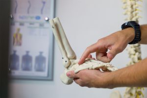
I am 40 years old. On June 21, 2019, I sprained my ankle. Since then, I’ve undergone Emet, Bowen, osteopathy, radiotherapy, and various forms of physiotherapy and kinesitherapy, but the swelling in my ankle has not subsided. I am unable to wear shoes, and walking is quite painful. An MRI revealed a tendon rupture and damaged ligaments. I have muscle dystonia. I know that without an examination, you can’t do much, but can you at least recommend some exercises or similar treatments?
If the MRI did not show cartilage damage, there must be a reason for the swelling (which is just a defensive, or protective mechanism). In your case, it is most likely ankle instability (ruptured ligaments, tendons, and dystonia combined). This means that while walking, the ankle and upper foot may be slightly “slipping,” and swelling is the body’s way of increasing pressure in the joint to reduce this “slipping.” If this hypothesis is correct, it explains why pain-relieving therapies (Emet, Bowen) were ineffective. It also explains why tension redistribution therapies (Osteopathy) didn’t help, and why treatments for local inflammation didn’t improve the situation (physiotherapy, radiofrequency therapy).
Additionally, I assume the exercises you’ve done were mainly focused on the foot and lower leg, which doesn’t have the potential to solve the issue in your case, as local muscle strength in the foot is not the problem here.
There are three possible solutions.
The first is to simply allow time to heal the problem. In a small number of patients, the body stabilizes the joint over time, either by changing the ankle’s anatomy, the movement patterns, or both. Surgical ankle stabilization (ligament reconstruction) is worth considering and discussing with an orthopedic surgeon. The third option is a complex exercise program aimed at re-stabilizing the ankle through the reactivation of the entire leg and torso because walking only ends with the foot but does not start with it. Our experience with this approach shows that persistent patients can achieve significant relief, and in many cases, complete elimination of symptoms.
I need advice regarding my problem.
Namely, due to large breasts, my spine hurts.
I know I need to strengthen my core, but would some relief come from wearing a posture correction belt for 3 hours – the one worn for clavicle fractures sold by Bauerfeind?
Every, and I mean every restriction of movement (it doesn’t have to be full immobilization) weakens the muscles. Braces used for clavicle fractures are no exception. They position the shoulders in their posterior position, aiming to align the clavicle fragments in a relationship from which we expect faster healing.
I understand how such support may seem attractive to patients with problems similar to yours, but by bringing the shoulder blades closer together, it further weakens the interscapular muscles and shortens them, worsening the existing painful condition.The same happens with all other elastic and inelastic braces and supports offered as a solution for either pain or posture. Initially, they seem to help, but after some time, the symptoms inevitably worsen.
The movement of the human body is a complex process developed through evolutionary “alchemy” aimed at reducing energy expenditure in movement. In this sense, it’s not only important to increase the strength of the core muscles but also their flexibility, so that through their elasticity, they reduce stress on the surrounding tissues and thus decrease the effort of maintaining a static position. Then, aerobic training (cycling, running, elliptical trainer, stepper) should help eliminate some of the consequences of long sitting or standing, for which evolution has not prepared us well. Next, exercises for the extremities should eliminate another part of the damage caused by prolonged static posture and strengthen the helper muscles of the spine. All of this should be done regularly (at least three times a week) as therapy and then as prevention of recurrence of symptoms.
The body is plastic. After some time, it will adapt to what we demand from it. If you minimize your movement, everything will weaken and atrophy, resulting in unpleasant symptoms. The remedy is simple. Do the opposite.
I have already undergone surgery on my right knee twice. This happened after 10 years of engaging in sports.The first operation was for the patella (dislocated patella) which was relocated.. after six months.The second operation was for the meniscus and cartilage.
Despite all this, I still experience pain. The exercises for the quadriceps are too painful, so I no longer do them. My orthopedist says this is normal. I haven’t engaged in any sports activities since the first operation and I don’t perform heavy physical tasks. I have had an MRI done.
Conclusion:
Signs of patellar cartilage chondromalacia (Grade I according to Outerbridge)
Chondral lesion of the medial articulating surface of the patellar cartilage
High patella (patella alta)
What does the MRI indicate, and what should I do because I no longer know how to help myself?
Generally, knee pain due to cartilage damage, especially on the patella, requires a more complex approach to treatment, particularly in terms of exercise, beyond just strengthening the muscles.
Namely, there is a reflex at work that, when knee pain is present, directly reduces the maximum strength of the quadriceps muscle through the spinal cord. This happens because the body, with reduced strength, attempts to reduce the force acting on the damaged cartilage. However, exercising in pain exacerbates this reflex and directly weakens the muscle, contrary to the desired outcome. If we don’t exercise, the muscle will weaken even without this reflex. Additionally, reduced movement impairs cartilage metabolism, making it more sensitive to pressure, which increases pain and further weakens the muscle. It’s a vicious cycle.
This situation can be remedied by combining two types of knee and surrounding muscle activation. The first is aerobic exercise, specifically riding a stationary bike with minimal resistance. Such movement accelerates cartilage metabolism and reduces its hypersensitivity. Then, you can start with the first pain-free and indirect, and then direct strengthening of the quadriceps, gradually intensifying the exercises.
Along this path, some methods of manual therapy can be beneficial (patella and knee mobilization/manipulation, trigger points, fascial manipulations). Isokinetic exercise can significantly shorten the recovery time as it minimizes pressure on the patella during muscle activation.

One of my late mentors used to say that the human body has a warranty period until 30 and a usable period until 40. Everything after that, in his opinion, is a bonus.
This statement, which he often shared with patients and students to raise awareness about preventing degenerative diseases of the musculoskeletal system, often hits right at the heart of the problem.
Evolutionarily speaking, this claim is entirely accurate. It’s only through advancements in medicine (particularly vaccines and antibiotics), hygiene, and food availability that human life expectancy has stretched to the current (expected) eight decades. What hasn’t changed is our genetic inheritance and the capacity of our metabolism to deal with the accumulation of minor damage to the musculoskeletal system, and then the consequences of such damage. Osteoarthritis, spondylosis, and other similar conditions, along with their impact on our daily lives through pain or limited function, are simply the final result of this damage.
For some patients seeking help for an acute condition based on degenerative changes (most commonly back and neck pain, but also knee, hip, ankle, and shoulder issues), the real question isn’t why the pain has appeared now, but why it didn’t appear until now. This shift in perspective allows us as professionals to see just how much the body can compensate for physical stress accumulated over the years, and it also gives patients an opportunity to reflect on how they treat their bodies, how much they invest in them (time, not money), whether they control their diet, and if they are regularly physically active. These are the key factors in preventing discomfort from degenerative changes, which we are all prone to—some luckier ones less so, the rest of us a bit more.
In this sense, the need for regular physical activity, adapted to age, physical condition, and personal preferences, only increases with age, rather than decreases. This is not only to reduce current symptoms but also to preserve flexibility, endurance, strength, and peripheral circulation. Mobility levels are directly correlated with the quality of life across all age groups.
MRI findings of the right ankle: chronic, minimal tendinosis of the proximal, partially involved part of the Achilles tendon. Intra-articular effusion and fluid collections adjacent to the lateral malleolus, minimal in the bursa of the Achilles tendon accompanied by tenosynovitis of the long peroneal muscle tendon, thickening of the plantar fascia, cellulitis of the soft tissue structures.
Despite months of physical exercises and spa treatments, the pain remains the same, with burning sensation in the area of the Achilles tendon even at rest, and it’s worst in the morning when getting out of bed. Extracorporeal shock wave therapy has been performed as well. Please advise me on the direction of my treatment, what do you suggest, are massages recommended, considering the inflammation of the tendon? Kind regards.
The MRI findings indicate “inflammation of everything,” from the Achilles tendon and plantar fascia to some other tendons in the foot, and even irritation of the ankle joint. In such a condition, conventional physiotherapy and shock waves are not helpful. With due caution, as I have not conducted a proper examination, it’s possible to interpret this finding as “tired foot,” meaning the foot is overloaded for a prolonged period beyond its capacity for recovery.
Whether in this particular case it’s due to excessive body weight, a predominantly standing or walking job, or a combination of these factors, along with insufficient physical activity and stretching, is less important. What’s important is to understand that massages will not be helpful. Some interventions in the field of manual medicine such as joint mobilization techniques, fascial mobilizations, or dry needling may provide some relief, but the solution to your problems lies in appropriate physical activity, prescribed by a physiotherapist after examination.
This includes aerobic exercises (stationary bike or elliptical training), strengthening exercises for the lower back and legs (as weak muscles transfer excessive load to the Achilles tendon), and intensive and proper stretching. Weight reduction, if excessive, is of great importance in such findings. As the tendon metabolism is slow, patient persistence in the treatment process is as important as the expertise of the physiotherapist leading it.
 In a lengthy letter, one of our readers describes issues with her chronic lower back pain. She is very physically active, attending high-intensity group workouts, which she wishes to continue. MRI scans show minor degenerative changes, and she asks if an exercise program could be developed for her based on the scans so that she does not have to come in for an examination. She lives in Zagreb.
There are things that, despite the best intentions, cannot be resolved via a mobile app or email. One of these is diagnostics in musculoskeletal medicine, and the other is therapy. Perhaps this option will exist in some indefinite future, but it is not available today.
Imagine you feel pain in your left heel while walking. You take off your shoe and see a blister. The first thing you will ask yourself is what caused the blister. Common causes are: a shoe that is too tight, a shoe that is too loose, laces tied too tightly or too loosely, bad socks… The same mental process occurs in the mind of an orthopedist or physiotherapist when making a diagnosis. In this case, MRI scans indicate where the “blister” is, or the location of the pain. Just as a simple photograph of the blister on the heel would indicate its location and size, an MRI shows the location and size of the pathological change. However, just as a photograph of a blister does not show the condition of the shoe, an MRI cannot indicate possible causes of these changes.
Degenerative and consequently inflammatory changes in the lumbar spine are, in most cases, the result of cumulative, gradual damage, resulting from overloading. The most common culprits are mobility disorders of the thoracic spine, hips, lower extremities (stiffness, shortened muscles, limited mobility), then localized or general muscle weakness, excessive or poorly directed physical activity, etc. None of these conditions can be read from MRI findings or other imaging diagnostics. This requires a proper and detailed examination, as well as a detailed medical history, in this specific case including the diagnosis of the training process, followed by individual therapy tailored to correct the identified deficiencies.
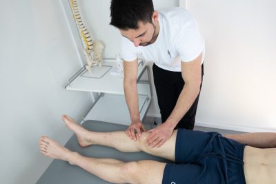
Seven days ago, I fell on the concrete, hitting my right knee and my left leg just below the knee. The next day, I felt as if there was swelling on that spot on my left leg, while my right knee was fine.
When I touch the area where my left leg was hit, it feels like the leg is numb, as if you’re running your hand over it, and there’s a stabbing pain when pressure is applied. I can move around freely, walk, and there are no problems until I touch the injured area.
The pain and “swelling” are the same intensity even after 7 days. There’s nothing visible on the surface, no sign of swelling, and no bruising. I don’t know what it is and what I should do, wait for it to pass or is there something for treatment?
The peripheral nervous system is composed of a network of nerves branching into smaller branches. This structure allows for the collection of sensory information from the entire surface of the skin, as well as from deep tissues. During trauma (strikes, twists, sprains, strains), nerves can be damaged along with other soft tissues and bones. In this case, it’s a minor nerve ending beneath the skin that’s damaged by the impact, likely accompanied by a small hematoma.
In a large percentage of similar situations, the sensation of numbness and increased tenderness upon pressure typically resolves on its own and doesn’t require any treatment. It may take weeks, or occasionally months, for full recovery. If you wish, you can expedite the process by gently massaging the area of discomfort and its surroundings for 5 minutes two to three times a day. It’s important that the massage is gentle. This will help with the absorption of any present hematoma and improve circulation around the damaged nerve, which should accelerate recovery. Such transient damage to peripheral nerves is called neuropraxia.
I’m interested in your opinion.
The cervical spine X-ray shows degenerative calcifications of the anterior longitudinal ligament at the C4/5 and C5/6 levels.
The thoracic spine X-rays distinctly show incipient ventral spondylitis.
The lumbar part is not good either.
What do you suggest for treatment?
The descriptions of the spinal X-rays in this case show moderate degenerative changes at the described segments, which can be common for a middle-aged person. No degenerative change is “good”; they represent the consequences of aging and lifestyle, much like wrinkles on the face.
Also, based on the description of the degenerative changes seen on the X-ray images, it is not possible to determine if the patient has any issues and what they might be. Therefore, determining treatment based solely on this information is not meaningful since these changes cannot be “treated” or otherwise influenced. In the end, it should be remembered that the descriptions of the images made by the radiologist are just that – descriptions of the differences in your images compared to an idealized image, which few people have after their 20s. The interpretation of these descriptions should typically be done by an orthopedic surgeon, physiatrist, and/or an experienced physiotherapist, based on a detailed physical examination.
That examination should provide information about the function of your spine (mobility, possible pain during specific tests, muscle strength and activation, condition of hips and shoulders, detailed medical history with frequency and type of symptoms). All of this is then put into the context of your daily activities, including physical ones if any, to conclude with an assessment of the current condition and a recommendation for actions to improve it. These procedures may include physiotherapy, changing some lifestyle habits, appropriate physical activity, or a combination thereof.
In short, degenerative processes are a combination of genetic predisposition and lifestyle. And while we cannot influence our genes (yet), our own habits are solely in our hands, making their change often the focus of conservative treatment.
Ten years ago, I fell while skiing and injured my knee, and since then my knee cap (patella) always dislocates. I’ve always had problems; if I stand awkwardly, it dislocates, and after so much time, my knee started hurting and swelling, and I have fluid in my knee.
The doctor at the Emergency Room looked at it with an ultrasound. Now I’m wondering what I need to do and if it’s normal for my knee to always dislocate.
Thank you in advance.
The symptom description indicates a luxating (unstable) patella. The medical history further suggests that trauma was the initial cause of this instability. The current condition, characterized by swelling and pain, indicates damage to other knee tissues (likely cartilage).
In cases of chronic instability of any joint (in this case, the knee, specifically the patella), over time, there is damage to other parts of the joint, with reactive changes, especially in bone, all in an attempt by the body to reduce instability. Gradually, and sometimes very quickly, this initiates degenerative changes leading to osteoarthritis, which is very difficult to treat. Because of this, joint instabilities that do not settle with persistent physiotherapy (especially exercises) should be surgically stabilized whenever possible.
Depending on the condition of the joint and the patella, it may be possible to stabilize the patella using a procedure called MPFL (medial patellofemoral ligament) reconstruction, which restores ligamentous stability. When performed by an experienced surgeon, this procedure typically resolves such instabilities, with a reduced risk of further osteodegenerative changes.
The postoperative recovery is relatively long, and it’s important to be guided by an experienced physiotherapist.
If there has been significant damage to the joint cartilage or other knee structures, then the mentioned procedure may not be possible, or a more extensive and complex surgical approach may be required. All details in this regard should be discussed with the surgeon, following a magnetic resonance imaging (MRI) scan, which should provide more detailed information about the condition of the knee.
About six months ago, I was diagnosed with a partial tear of the supraspinatus tendon.
An MRI examination and consultations (Lovran and Matulji) led to the recommendation of arthroscopy. I have decided to undergo the procedure in about 2-3 months, but I am interested in whether, in your expert opinion, it is better to wait passively or actively (engage in exercises with you).
I occasionally experience significant pain. I am 73 years old.
When deciding on the treatment of partial tears of the rotator cuff of the shoulder (in this case, the supraspinatus), several questions are important. How large is the tear, is it traumatic or degenerative, what is the age of the patient, and what are the demands the patient places on their shoulder (how physically active are they)?
In this regard, we typically aim to surgically repair most traumatic partial tears up to the age of 30 or 40 because the alternative is living with significant physical limitations, and we expect further damage to the shoulder. Unfortunately, rotator cuff tendons hardly or never heal on their own. After this age, especially concerning degenerative tears (which occur gradually as a result of use with poor regeneration), we assess what is best in the specific case.
Degenerative changes in tendons make them fragile, and the surgeon may have difficulty reducing or completely covering the tear. The postoperative recovery is long (at least 6 months) and requires complete dedication from the patient. Due to the condition of the tendons, even when ideally “sutured,” their healing is not guaranteed. Therefore, it is extremely important to undergo appropriate therapy before surgery to reduce pain and establish the best possible shoulder function in the current state. If the condition after therapy is satisfactory for the patient, then the surgeon will have an additional criterion to question the need for surgery. If the condition after therapy does not meet the patient’s requirements, then excellent preparation for surgical intervention has been made, and the prognosis for its outcome is better.
I am 40 years old. On June 21, 2019, I sprained my ankle.
So far, I have undergone Emet, Bowen therapy, osteopathy, radiotherapy, and various forms of physiotherapy and kinesiotherapy, but the swelling in the ankle is not reducing, so I cannot wear shoes, and it is quite painful when walking.
The MRI showed a tendon rupture and damaged ligaments. I have muscle dystonia. I know that without an examination, you cannot do much, but can you at least recommend exercises and the like?
If the MRI did not show any cartilage damage, there must be a reason for the swelling (it is merely a defensive or protective mechanism). In your case, it is most likely ankle instability (ligament rupture, tendon damage, and dystonia combined). This means that during walking, the ankle and the upper part of the foot slip slightly, and the swelling is a way for the body to increase pressure in the joint and reduce these slips.
If this hypothesis is correct, it explains why therapies aimed solely at reducing pain (Emet, Bowen) have not yielded results. It also explains why tension redistribution therapies have not been helpful (Osteopathy). And finally, it explains why treating local inflammation does not bring improvement (physiotherapy, radiofrequency therapy). Also, I assume that the exercises you have been doing have mainly focused on the foot and lower leg, which is an approach that does not have the potential to solve your problem because local muscle strength in the foot is not the issue in your case.
There are three possible solutions. The first is to simply let time heal the problem. In a small percentage of patients, over time, the body stabilizes the joint, either through changes in the anatomy of the ankle itself, changes in movement patterns, or a combination of both.
Surgical stabilization of the ankle (ligamentoplasty) is worth considering and consulting with an orthopedist.
The third option is a comprehensive exercise program aimed at restabilizing the ankle, but through the reactivation of the entire leg and trunk, as movement when walking does not only end with the foot but does not start there either. Our experience with this approach shows that in persistent patients, it has the potential to significantly reduce symptoms and often even make them disappear completely.
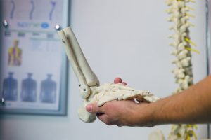
I’m interested in how many treatments are typically needed to treat a heel spur?
The size of the spur is 13mm/0.55inch.
A heel spur is a finding on X-rays. There are so-called “upper” and “lower” heel spurs. The first one appears at the attachment of the Achilles tendon to the heel bone. The second represents a bony protrusion in the lesser-known muscle, flexor digitorum brevis (short toe flexor). Above this protrusion lies the large ligament called the plantar fascia, whose attachment can become inflamed in a way that causes pain in the lower part of the heel during walking, and in acute conditions, even at rest. In this sense, the cause of the pain is not the bony protrusion (spur) but the inflammation of the plantar fascia attachment due to overuse. This overuse can be caused in various ways, such as flat feet, previous ankle sprains, prolonged standing or walking, especially in inadequate footwear, and similar factors. Based on the assessment of the probable cause of inflammation and its condition, therapy is determined, and its duration is estimated. Since we do not treat the spur itself, its size does not affect the method and duration of treatment. Treatment may only involve changing footwear or making orthopedic insoles. In addition, for some patients, stretching and proprioception exercises alone may be sufficient. For some, one or several manipulations of the small foot joints may be enough. In other cases, it is necessary to stimulate the healing of the damaged and inflamed ligament area through a combination of manual therapy and physical therapy modalities (laser, ultrasound, radiofrequency therapy, FMS…). In the most severe cases, when the condition requires it, shock wave therapy (ESWT) can be applied. The duration of treatment depends on the specific case and therapeutic approach and can be approximately determined at the first examination, during which we assess the level of inflammation and damage to the plantar fascia using ultrasound diagnostics.
“Hello, I am 27 years old and I have hip arthritis.
I am a bit too young for surgery, so I’m wondering if there is any fluid that can be injected into the hip joint because I’m constantly in pain.
Cartilage regeneration or something… thank you.”
Arthritis in the hip at such a young age is usually the result of congenital deformity or trauma. Regardless of the initial cause, therapeutic protocols are similar and consist of three main parts.
The first is analgesic therapy, which can be done with traditional physiotherapy (laser, ultrasound, magnet, currents), but our experience shows that these methods are rarely successful. On the contrary, by combining techniques of manual therapy (mobilization and manipulation techniques, trigger point stimulation, fascial manipulation) with appropriate exercises and possibly radiofrequency therapy, we achieve a significantly better outcome.
After reducing pain, it is important to continue with basic exercises and gradually intensify and expand the program, aiming to maintain reduced pain levels or become pain-free.
Avoiding physical activities that further damage the cartilage is an important part of the rehabilitation process, especially when it comes to running, jumping, and sudden changes in direction.
Surgical procedures such as arthroscopy, “cleaning,” and “smoothing” of the cartilage, which are effective for the knee, are almost entirely useless for the hip and are rarely performed.
Unfortunately, a similar situation exists with injections into the joint. Hyaluronic acid, PRP, and the like have also not shown significant effects on hip arthritis. In this sense, if you wish, you can try instillations, but do not expect much from them. On the contrary, the basis of the entire recovery process is analgesic therapy combined with exercise.
It is worth noting that in this case, exercise not only aims to strengthen muscles and improve the flexibility of the joint and surrounding structures but also, by including aerobic “training,” the additional goal is to “condition” the remaining cartilage to become more resistant to physical stress. Based on this, we expect a reduction in pain through regular exercise.
Kada je, prije nekoliko mjeseci, jedan mladi nogometaš zatražio pomoć poradi bolova u lijevom koljenu, sve je ukazivalo da će liječenje biti relativno kratko i uspješno. Pregled i ultrazvučna dijagnostika ukazivali su na iritaciju hvatišta velikog butnog mišića na patelu, tzv. „skakačko koljeno“ uzrokovanog stanjem hrskavice na samoj pateli, te potaknutog intenzivnim treningom. Uobičajeno, terapija i odgovarajući trening trebali bi ukloniti simptome unutar nekoliko tjedana.
Kada se to nije dogodilo, proširili smo pretragu.
Cijeli lokomotorni sustav radio je bez problema. Pokretljivost kukova, stopala, zdjelice i leđa bila je uredna. Snaga mišića primjerena. Jedino što je bilo uočljivo, bio je ožiljak od operacije preponske kile dvije godine ranije.
Slijedeći principe modernih kliničkih anatoma Vleeminga i Stecco-a postavili smo hipotezu da je taj ožiljak, koji se pregledom pokazao slabije mobilan, pokrenuo „tenzijsku kaskadu“ koja se u konačnici pokazala kao bolnost u koljenu poradi specifičnosti same sportske aktivnosti.
Terapija se sastojala u mobilizaciji samog ožiljka, kao i u nastojanju smanjenja tenzije postupcima manualne terapije unutar cijelog „tezijskog lanca“. Zanimljivo u ovom slučaju je da je bilo potrebno tek dvije terapije za potpunu sanaciju simptoma. Prije nekoliko dana učinio sam i kontrolni pregled. Sada već bivši pacijent u punom je treningu i igra utakmice bez smetnji.
Svi mi koji smo educirani unutar klasičnog ortopedskog pristupa unutar kojega je uzrok smetnji na sustavu za pokretanje gotovo uvijek na mjestu ili u neposrednoj blizini boli (osim u slučaju pritiska na živac) imamo problem sa prihvaćanjem drugačijih ideja unutar kojih se tenzija može širiti vezivnim strukturama u ljudskom tijelu, te bol izazvati na udaljenim mjestima. Srećom, klinički rezultati novog vala razmišljanja daju nam dodatne mogućnosti dijagnostike, a potom i terapije, naročito u sindroma prenaprezanja, poput skakačkog koljena, teniskog lakta, petnog trna i mnogih drugih unutar kojih se klasičan fizioterapijski pristup ne može pohvaliti dobrim rezultatima.
So, I’ve been involved in athletics for about ten years now, currently I’m 20 years old. This year I underwent surgery on both knees. I had arthroscopy for synovial plica reduction and Hoffa’s fat pad reduction, diagnosed with patellar tendinitis and ACL issues.
The surgeries were in March and late May. I’ve completed physical therapy and exercise daily following the plan from DNS experts in Rijeka, but I still have pain.
My goal is to return to running as soon as possible because of my sports scholarship. Any advice? Thank you!
Anterior knee pain in running and jumping disciplines is a specific problem that has been the subject of numerous scientific papers and books. Our profession seeks to understand the causes of such issues, which are not always simple and localized only to the knee.
Namely, anterior knee pain in athletes is always the result of overuse of tissues in the knee extensor mechanism. We understand that intensive training is usually the cause of this overuse. However, the same training will not cause overuse in all athletes. This means that individual differences in various aspects of body form and function can contribute to the development of inflammation and pain. Thus, besides the condition of the knee itself, some specific characteristics of the hip, foot, and spine can create an environment where stress from training is not evenly distributed but rather focused on the knee, causing pain.
The surgery, I believe, addressed the local problem. In this case, the internal folds on the joint capsule and the fatty cushion called Hoffa’s body. It is possible that residual hip stiffness, altered spine kinematics, or some other of many possible conditions, which do not hurt by themselves but increase stress during running. It is also possible that postoperative scars, both internal and external, stimulate pain receptors and thus support pain.
I recommend seeing an experienced sports physiotherapist. They should be able to assess which of the possible issues are present in your case, advise you on how to address them, and thus alleviate the pain.
I am 33 years old. About a year and a half ago, while training in the gym, I felt pain in the left side of my back while doing squats and deadlifts. After a few days, the pain didn’t completely go away, but I thought it would disappear like everything else, and I would be able to train again. However, when I tried to train again, after a very short time, the pain returned fully. The pain occurs only in the left side of the back, never in the right, and has been present more or less constantly for a year and a half now. The pain most often appears when rotating to the right and bending forward. I don’t feel pain in the spine, only in the muscle, which I’m unsure if it’s strained or tight. I’ve had several massages and therapies, but no one has explained the cause of the pain to me, nor has it ultimately helped.
Most lower back pains resolve spontaneously and without treatment within a few days to a few weeks from onset. Of those that do not subside, about half will respond well to non-specific therapies such as massages, some physiotherapy procedures, or manual/manipulative techniques. If this does not happen, it is time for diagnostics. This starts with an examination by a specialist doctor, an orthopedic surgeon, a vertebrologist subspecialist, or a neurosurgeon, and in exceptional cases, a very experienced physiotherapist can perform it. The goal of this examination is to make a working diagnosis or hypothesis, which is the basis for further diagnostics. Based on your description of symptoms, it is reasonable to assume that you may have a disc deformation pressing on the surrounding soft tissues. As this deformation can be directed more to the left or right, the discomfort can be localized to only one side of the back. If the deformation is not severe, then the symptom is a defensive muscle spasm. If this assumption is correct, which needs to be confirmed by examination, an MRI of the spine would be recommended. Based on the MRI findings, a suitable treatment program is created, along with advice on physical activity that won’t further provoke the discomfort.

