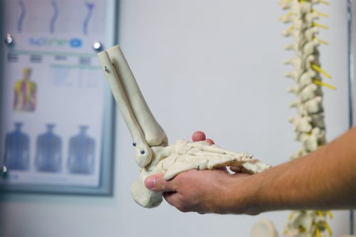
The plantar fascia is actually a broad ligament that extends along the underside of the foot, from the heel bone all the way to the toes. It has three functions, two of which are well described in the literature.
Plantar fasciitis
Description
One is maintaining the arches of the foot. The second is functioning as an extension of the Achilles tendon, through which the forces of the calf muscles are transferred to the toes. The third, less studied and known, is its role in the foot’s shock absorption system, responsible for absorbing stress during walking, standing, running, and other athletic activities.
Plantar fasciitis, or plantar fasciopathy, refers to a condition characterized by pain in the heel and its surrounding area, which is most intense in the morning after rest, decreases after the first few steps or hours, and then worsens again throughout the day, especially after prolonged standing, walking, or physical activity.
In plantar fasciitis, the attachment site of the plantar fascia to the heel bone or the part near this attachment becomes thickened, its structure changes, and it becomes inflamed as a result of overstrain. The inflammation here, as in other overuse syndromes, is not the classic type that leads to healing, but rather a chronic condition where the inflammatory process stops halfway, not progressing toward healing, resulting in persistent pain that does not subside with rest or over time.
Depending on a range of factors, heel pain may only be present in the morning after rest or prolonged sitting, and it may vary from mild to moderate or sharp and shooting. It may also worsen with standing or walking, but lessen with rest, or it may be constant, with occasional flare-ups.
This overuse syndrome occurs in athletes, especially in sports dominated by running or jumping, but it is relatively rare. It most commonly appears in middle-aged and older individuals, especially those who stand or walk a lot throughout the day. Literature suggests that age-related changes in the connective tissue and the natural flattening of the foot arches over time are contributing factors. In clinical practice, risk factors associated with its onset include flat feet or high arches, previous ankle sprains, increased body weight, relative or absolute physical inactivity, sporadic physical activity with intense phases, wearing inappropriate work or sports shoes, hormonal imbalances, and certain systemic diseases.

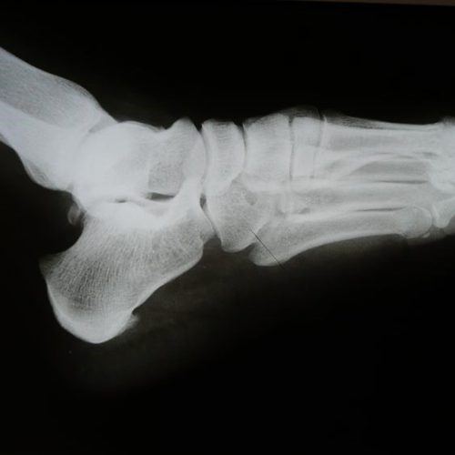
Diagnosis was until recently relatively simple and in most cases involved just a specialist’s examination. Specific pressure pain, as well as pain during movement (walking on toes and heels) and stretching, clearly indicate this syndrome. X-rays may also show the presence of a “heel spur“, which is a bony outgrowth in the little-known flexor digitorum brevis muscle (a short flexor of the toes), located beneath the plantar fascia itself. However, there is no direct connection between pain and the presence of a heel spur. I would argue that there is no connection at all. There are enthesophytes (bony growths) that are not associated with pain, as well as very painful plantar fasciitis cases without changes in the heel bone. In most cases, physiotherapy, along with appropriate support and changes in daily activities, can reduce or eliminate the pain, even if the “heel spur” remains. Therefore, the visibility of a “heel spur” on X-rays has no diagnostic value, yet orthopedics, orthopedic physiotherapy, and consequently patients, often believe that this bony outgrowth is the source of all their problems, which can present special challenges in clinical practice.
Ultrasound diagnostics, routinely performed in our clinic as part of the initial examination, show that changes in the shape of the plantar fascia and its structure, along with the inflammatory process, are what cause the pain. Additionally, this method allows us to rule out other potential causes of heel pain, such as a rupture of the heel fat pad, plantar fibromatosis, or a rupture of the plantar fascia itself, which are relatively rare conditions but present with the same symptoms as plantar fasciopathy, though their treatment is significantly different.
Treatment, as with all overuse syndromes, can be divided into local and global approaches. Local therapy has two possible directions. One is a combination of manual therapy for the joints and soft tissues with modern technologies available in physiotherapy, such as radiofrequency therapy, laser, or FMS. The goal of this approach is to calm the inflammation through direct stimulation of biological processes, along with mechanical stimuli to alter the physical characteristics of the plantar fascia and the surrounding ligamentous system. If this approach does not yield satisfactory results, or if during the diagnostic process we assess that it is unlikely to help in the specific case, we apply shockwave therapy (TUV-ESWT), where sound waves stimulate the formation of new inflammation, which, through its own cascade reaction, should lead to the healing of the damaged tissue by forming scar tissue, thereby eliminating the existing symptoms. Each of these therapies is successful in about 70% of cases, and together in about 90%.
Tension Model
The relationship between the plantar fascia and the bones and small joints of the foot can be likened to the relationship in a bow used to shoot arrows, between the bow itself and the string that draws it. In this example, the string is typically inelastic, as is the case with the plantar fascia, and the bow is elastic, although the bones and joints of the foot are not naturally elastic on their own. Their elasticity is achieved through small movements that the foot joints make with each step. This allows for excellent shock absorption during walking, running, and other sports and physical activities. A reduction in these movements, through restricted mobility of one or more joints, stiff tendons or ligaments, or changes in foot kinematics after sprains or other traumas, also reduces the foot’s shock absorption potential. This diminished shock absorption directly affects the plantar fascia by increasing the amount of force it is subjected to along its length. Over time, these forces produce changes at the attachment point or along the course of the plantar fascia, leading to thickening and, later, a chronic inflammatory process known as plantar fasciitis or plantar fasciopathy. Understanding this mechanism makes it clear that one of the key treatment goals is restoring proper shock absorption through manual therapy techniques, specific exercises, and, when necessary, the use of appropriate orthopedic insoles.
However, shock absorption during movement involves the entire lower extremity, of which the plantar fascia is just a small, but important, part. Limitations and reduced shock absorption capacity in structures around the lower back, hip, knee, or ankle can be directly transferred to the plantar fascia as increased tensile forces, causing the pathological changes and symptoms described earlier. Therefore, diagnosing distant causes in the large kinetic chain is an important part of the diagnostic process, and addressing them is an essential part of successful plantar fasciitis therapy.
All of the above is part of the standard individualized treatment protocol for this painful syndrome at our facility.
A few additional pieces of information and advice:
-Creams, ointments, and gels have no effect on plantar fasciitis. The thickness of the skin and subcutaneous tissue prevents any substance from reaching the plantar fascia.
-Shockwave therapy cannot “break” the heel spur. It represents a bone shape change, and shockwaves have no effect on its size or shape, regardless of what “information” you find on the web or social media. On the contrary, this method is highly successful in treating pathological changes in soft tissues like tendons and ligaments.
-Heel lifts and silicone pads are typically effective in reducing pain, but only temporarily. The pain usually returns to its original intensity after a few days or weeks.
-A “cortisone and anesthetic block” can be administered to the site of inflammation. The short-term results are good, but in most cases, the pain returns after a few weeks or months, requiring either another block or appropriate physiotherapy. For this reason, and due to the increased risk of infection with injections into the foot, as well as the fact that cortisone, which reduces inflammation, also “weakens” the connective tissue and can significantly damage the fat pad, turning a minor painful syndrome into a serious condition requiring complicated and lengthy treatment, we do not use this method at our facility.
–Surgical procedures for plantar fasciitis are possible but are performed very rarely, as conservative methods are generally successful, and the final results of surgery are rarely truly satisfactory.


