Ankle sprains are the most common injury in that part of the body. Sprains can be classified by severity into mild, moderate, and severe, and by type into inversion (twisting inward), eversion (twisting outward), and plantar, where the entire foot is bent backward.
Description
Americans have calculated that approximately one million of their citizens seek medical attention each year for this injury, which, translated to Croatian figures, suggests around 16,000 patients with ankle sprains in doctors’ offices. Additionally, statistics indicate that between 10 and 30% of all sports injuries are related to sprains, and perhaps most importantly, up to 40% of all sprains risk developing into chronic problems in the ankle or surrounding areas. Inversion sprains are by far the most common, eversion sprains are much rarer, and plantar sprains are very rare.
Assessing the severity of a sprain is difficult immediately after the injury. It is typically estimated based on the size of the swelling, difficulty or inability to walk, and the level of pain. However, the size of the swelling depends more on the size of the blood vessel damaged during the sprain, and consequently the amount of blood that has leaked into the subcutaneous, intra-articular, or interstitial space, than on the extent of tissue damage such as ligaments, tendons, or joint cartilage, which are the real indicators of the injury’s severity. Furthermore, the size of the swelling depends on the number of previous sprains, where each subsequent sprain typically causes less swelling but may involve greater damage to the aforementioned tissues.
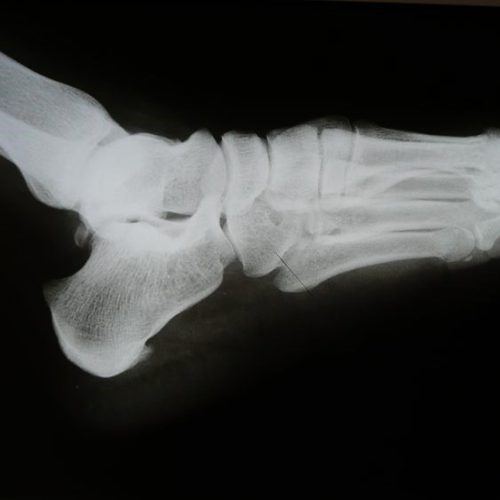
X-rays can rule out the possibility of bone fractures. Ultrasound can assess the size and location of the swelling and the extent of damage to some, but not all, soft tissues. MRI provides the most detailed view of the damage but is rarely prescribed immediately after an injury due to its limited availability and cost. However, even all the details it can provide may not be crucial in estimating the duration of treatment and recovery. This is because recovery speed depends not only on the size and type of tissue damage but also on individual characteristics that can only be assessed through a detailed physical examination. It is also important to emphasize that ankle sprains are very rarely localized only to the ankle. They almost always involve injuries to the small joints of the foot and surrounding tendons, all of which influence the assessment of the injury’s severity, the duration of treatment and recovery, potential long-term consequences, and the speed of return to sports, if the person is physically active or an athlete.
The good news is that at least half of all ankle sprains do not require treatment and heal spontaneously with some rest and avoidance of physical exertion. The problem lies with the other half.
One common issue is immobilization (with a splint, cast, or brace) and its duration, which remains a point of disagreement among professionals. Namely, ligaments injured during a sprain do not heal better if the foot is kept immobilized; therefore, the purpose of immobilization is to prevent further damage and provide the freshly injured tissues with a calm environment to promote initial or primary healing. For this reason, our institution recommends immobilization after an ankle sprain, but for no longer than 10 days post-injury. We consider anything longer to be counterproductive, as it prolongs treatment time and may even lead to biological processes that ultimately hinder complete healing.
The protocol we recommend involves an examination (by an orthopedist or experienced physiotherapist) and an X-ray to rule out bone damage.
Immediately after the sprain, an ice pack should be applied. The best way is to prepare ice cubes in a plastic bag and place it directly on the painful area. After ten minutes, the ice pack should be removed, and the injured area should be wrapped with an elastic bandage. This is done to reduce swelling, which in turn speeds up the overall recovery process. The purpose of the ice is vasoconstriction (narrowing of the blood vessels), while the elastic bandage increases surrounding pressure on the injured blood vessel to promote earlier cessation of bleeding. Although there are various approaches and opinions in the field about how long ice should be kept on the injured area, it is advisable not to overdo it. In my practice, I have seen many cases of frostbite that ultimately prolonged recovery rather than shortened it. Similarly, I do not recommend applying other frozen items (such as frozen meat, peas, etc.) directly to the painful area for the same reason. If ice is not immediately available, cold tap water can help. After the elastic bandage is applied, ice can be placed over it. At bedtime, the bandage should be removed to allow circulation of blood and lymph. Research has shown that cold and ice packs help in the first 2–5 days post-injury, but beyond that, they slow down healing and are not recommended as long-term therapy. The same goes for the elastic bandage, which loses its function after a few days.
If the X-ray shows that the bones are intact (no fractures) and there are no signs of a tear in the tibiofibular syndesmosis (a strong internal ligament that holds the two bones of the lower leg together), the recovery process begins. In the first 48–72 hours, recovery consists of rest, ice packs, elastic bandaging, and elevating the injured leg.
After a brief period of rest, physiotherapy begins in full, with the goal of reducing swelling and stimulating healing. Typically, during this phase, the area around the joint, but often the entire foot and a good portion of the lower leg, will take on various colors, from dark blue-black to yellow-brown, all as a result of hematoma resorption. Depending on the severity of the injury and the individual’s specific capabilities, the goal is to restore normal range of motion in the joint and bear full body weight on it as soon as possible. This phase of treatment can last from a few days to several weeks, depending on the size and severity of the injury. After this, recovery continues with exercises aimed at local and global stabilization of the ankle, which is the most important part of the overall treatment. It also includes preparation for returning to sports, when needed, with very specific procedures that vary from sport to sport (“sport-specific”).
In the vast majority of sprains, full joint function and that of all associated structures are restored by the end of treatment, allowing the patient to engage in daily and sports activities without problems. In a small percentage of cases, instability remains, presenting as occasional minor sprains during walking or physical activity, a sense of weakness, fear of sudden changes in direction, or, in later stages, the development of pain associated with anterior ankle impingement. All of this indicates manifest ankle instability and requires either the use of a protective brace during sports activities or surgical stabilization of the ankle, which involves the reconstruction of one or more damaged ligaments. These are generally procedures with good outcomes and relatively quick postoperative recovery.
Tension Model
There are two aspects of the tension model we use in the treatment of ankle sprains.
The first is the premise that damaged tissues under tension heal slowly or not at all. This is similar to a situation where you cut yourself and the wound is not closed (by stitches or bandages). Such an open wound under tension heals more slowly or not at all. The same is true for damaged tissues in the ankle and foot. It is important to reduce their tension after the injury. This is achieved through specific mobilization and manipulation techniques and methods of “taping” with rigid tape or adhesive bandages.
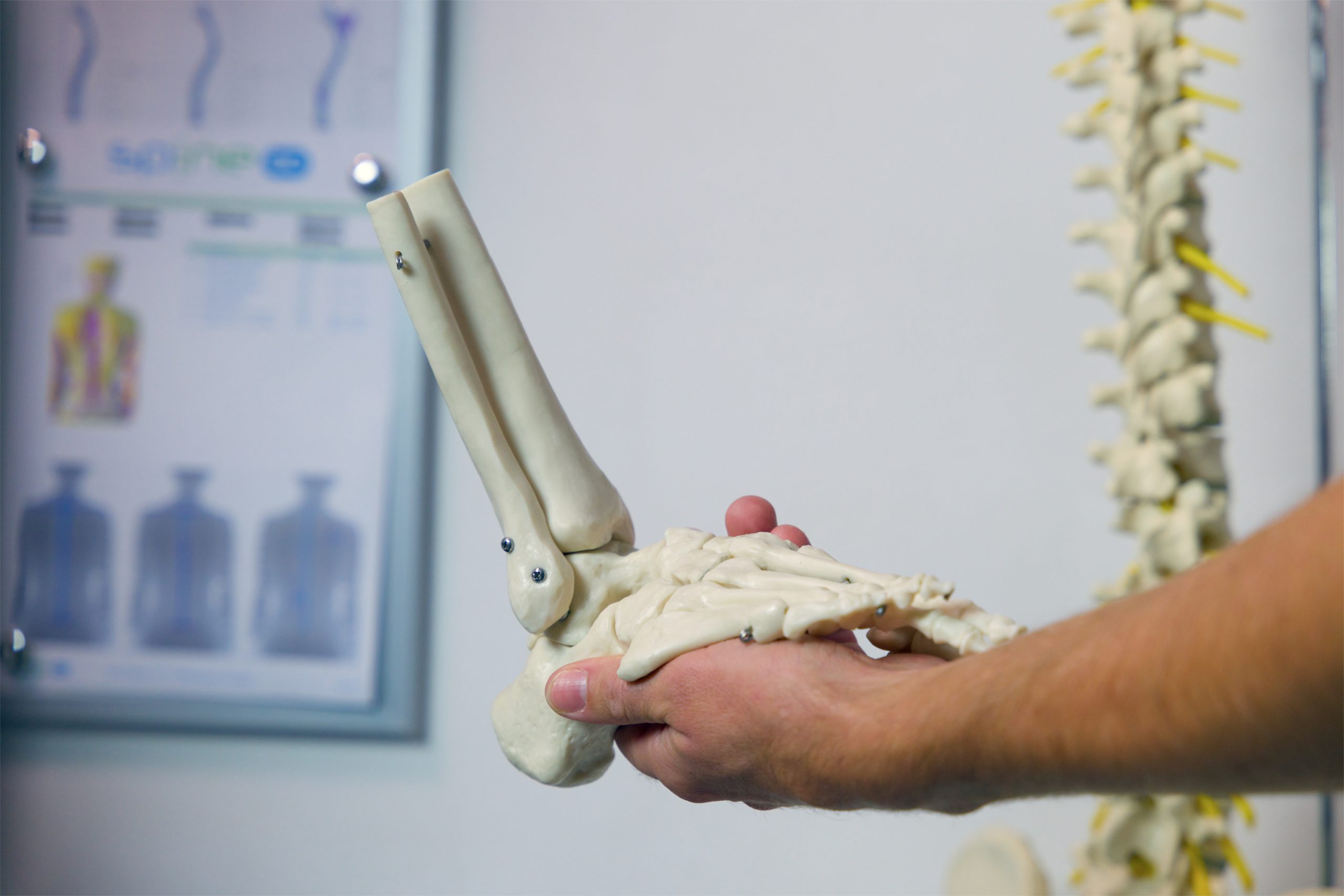
The second is an understanding of the anatomy and biomechanics of the foot, ankle, and entire lower limb as a functional unit. The muscles and tendons of the foot itself are relatively small and weak, and cannot stabilize the ankle alone, even when it is not injured. Ankle stabilization is the function of the entire lower limb, all its muscles, and their quick and timely activation. Global stabilization in large kinetic chains is the key to full recovery, especially in more severe sprains. In this context, the tension model provides detailed guidelines regarding the type, scope, and intensity of interventions needed to establish complete stability after injury.
A Few Final Tips:
- Manipulation (“realignment”) of joints is an extremely powerful intervention that can reduce pain, accelerate healing, and significantly improve overall treatment outcomes, but only when done at the right time, in precisely selected places, and by an experienced physiotherapist/manual therapist. I regularly encounter the consequences of poorly performed manipulations, and their remediation is part of my daily work. I wish there were less of it.
- “Breaking down” a hematoma through very aggressive massage is a popular procedure in sports settings. I believe that it is generally ineffective and can be harmful because it essentially involves inflicting new trauma on already traumatized tissue. It is especially contraindicated in the first 72 hours after the injury, as it inevitably leads to new bleeding and greater swelling. There are exceptions, but they are extremely rare, where this procedure may be helpful. Then, the experienced physiotherapist needs to make a careful assessment of whether the inevitable damage caused by this rough treatment will be less than the benefits we expect from its application.
- Ointments, creams, gels, and homemade remedies like vinegar or egg white compresses, objectively cannot help. If you insist on using them, do so with caution regarding the ingredients that are harsh on the skin and may cause irritation or allergic reactions. These consequences hinder or prevent the application of proven physiotherapy interventions that help, thus prolonging treatment.
- Any pain, swelling, or feeling of instability that persists six weeks after an ankle sprain requires an examination by an orthopedist and/or experienced physiotherapist.