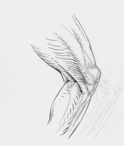
A small bone embedded in the tendon of the large thigh muscle, which we call the patella or kneecap, is colloquially and incorrectly referred to as the “cap.” It is located on the front side of the knee and is very easy to locate when you run your hand over that spot. Its primary role is to serve as a fulcrum for the large thigh muscle during knee extension, just as a seesaw’s pivot point is in its middle.
Patellar Chondropathy (Chondropathia, Chondromalacia)
Description
Unlike most bones in the body, the patella is a floating bone. Embedded in the tendon of the large thigh muscle, its back side is covered with cartilage, allowing it to glide during movement over the lower part of the femur. On X-ray images, the patella appears as a wedge, while the femur has a corresponding groove ensuring the patella’s movement path is always consistent. Essentially, it is part of the quadriceps tendon.
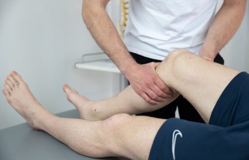
Patellar chondropathy is one of the most commonly diagnosed painful knee conditions across all age groups. It is characterized by pain at the front of the knee, which intensifies when walking, especially downhill or down stairs, and is present after rising from a seated position or during prolonged sitting. During an examination, pain is provoked by mobilizing the patella (its passive movement by the physician or therapist’s hands) and other compression tests. Squatting is usually painful. If an MRI is performed, some patients may show changes in the cartilage of the patella or the cartilage of the femur, over which the patella glides during movement. In such cases, chondropathy, a clinical diagnosis, is usually renamed chondromalacia, a radiological finding implying confirmed cartilage softening or damage.
Standard Treatment
Treatment is based on a range of physical therapy methods (electrotherapy, ultrasound, laser, radiofrequency therapy, magnetotherapy…), along with strengthening exercises, especially for the quadriceps. In particularly persistent painful conditions unresponsive to these methods, arthroscopy may be performed. This minimally invasive surgical procedure involves making two or three small incisions in the knee and using specialized instruments to smooth any irregularities in the cartilage or shave the thin, softened superficial layer of the same cartilage. The idea behind such procedures is that the treated tissue will cause less pain during movement. Unfortunately, the results of these procedures often fall short of the expectations of both the operator and the patient.
It is also possible to try hyaluronic acid injections to improve patellar glide during movement, a procedure that is increasingly used, with variable results.
The Central Problem
The fundamental issue with all these described standard interventions for patellar chondropathy is the premise that cartilage changes cause pain. However, cartilage lacks pain receptors or any sensory receptors, just as fingernails are not painful when cut. Thus, the question lingering over patellar chondropathy for many decades remains unresolved: What exactly causes pain?
Since cartilage cannot be the cause, based on basic physiological facts, what is happening?
 Potential Pain Sources
There are three possible answers, as there are three types of tissues in the knee that have pain receptors and nerve endings, with the potential to transmit pain impulses:
- Bone
- Synovial membrane (the inner lining of the joint, responsible, among other things, for joint lubrication)
- Fat pads (Hoffa’s, suprapatellar, and infrapatellar), whose role is to absorb forces during knee and patella movement.
Bone is painful when traumatized, reacting with inflammation or fracture. As this condition does not involve trauma, fractures can be excluded, leaving inflammation. This appears on MRI as a bright area in the bone beneath the cartilage. However, this condition is only present when cartilage is severely damaged, penetrating to the bone. Such situations are not uncommon but account for a very small percentage of all chondropathies, meaning they cannot explain the pain in most patients.
Irritation of the synovial membrane can occur as a result of a systemic disease such as rheumatoid arthritis or entrapment of part of the membrane during movement (like a synovial plica). Most chondropathies do not involve rheumatoid arthritis, and the symptoms of a synovial plica differ from those of chondropathy.
Irritation and excessive stress on fat pads is the last of the proposed options and carries particular weight because these tissues are very well supplied with sensors and nerves. Moreover, using newer and more sensitive MRI machines, small inflammations in these structures can be detected, which are visible in some, but not all, chondropathies, and not even in the majority. Furthermore, irritated and inflamed fat pads should be painful to the touch, which they are not in most patellar chondropathies.
To add further complication (for those who have followed the story thus far), this condition is more common in young girls than boys, particularly those involved in sports during periods of rapid growth. This has been attributed to the role of estrogen, which during the growth and development of girls acts as a stimulator of ligament flexibility. Since cartilage is derived from connective tissue during fetal development, it retains estrogen receptors and also becomes softened during rapid growth phases in adolescence. However, as we have seen, cartilage itself is not painful, so its softening, regardless of the cause, should not inherently be painful.
Moreover, during the development of patellofemoral arthrosis, which refers to the complete destruction of cartilage and changes in bone shape, some patients exhibit no symptoms even when there is little or no cartilage left.
There are more such examples, but they would only complicate matters further while pointing to the same issue and question: Where does the pain in patellar chondropathy/chondromalacia come from? We don’t know. Not only do I not know (I am just an insignificant physiotherapist in a clinic in Zamet, a small town called Rijeka, and I don’t know many things), but the medical profession also does not know.
This lack of knowledge leads to generalized therapeutic solutions, partly described in the text above, which help some patients but are entirely ineffective for others, possibly the majority.
In short, this is an example where we treat pain whose origin we do not understand with therapeutic interventions whose mechanisms of action in the specific case we also do not fully understand.
The tension model (click for article) developed in our clinic provides a hypothesis for the possible cause of pain in patellar chondropathies. It postulates that any prolonged increase in tension within the connective system causes pain because the connective system is richly supplied with mechanical and pain receptors. Similar to when any muscle or tendon is stretched long enough and strongly enough, the stretching will eventually turn into pain.
Similarly, the knee extensor system, consisting of the quadriceps tendon, patella, and the complex network of surrounding ligaments and connective structures, can become overly tense, either entirely or in part, for various possible reasons, causing direct pain or indirectly irritating the synovial membrane or fat pads.
This model predicts that reducing excessive tension would immediately reduce pain. This is precisely what we observe in patients with chondropathy during manual interventions aimed at achieving just that. These interventions serve therapeutic purposes but, above all, confirm the hypothesis. When confirmed, specific tests and examinations identify structures more tensioned than others, and potential local causes of this tension are explored (weak muscles, slow proprioceptive reactions, stiff adjacent joints like the hip or ankle, and a range of other mechanical and kinematic dysfunctions).
Interventions to address identified deficiencies follow, which, if properly applied, lead to satisfactory therapeutic results within a reasonable time for most of our patients.
Although this model, its hypothesis, and diagnostic and therapeutic procedures are not scientifically proven, they are based on only slightly different interpretations of known and scientifically proven anatomical and physiological facts and processes. They enable us to provide individualized and specific diagnostics and therapy, with significantly improved predictability of therapeutic intervention success and treatment duration.


