Imaging diagnostics (MRI, X-ray, ultrasound, CT…) is of great assistance in diagnosis, but when taken for granted, it can also be a major obstacle, as shown by the following example from our practice.
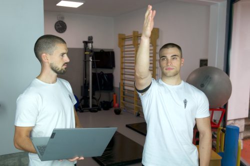
Neck and Shoulder
Description
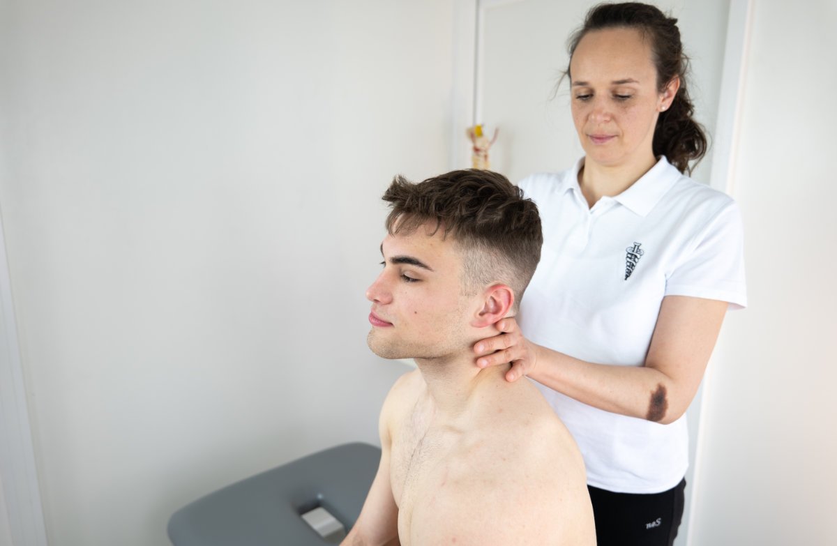
A few weeks ago, a 48-year-old woman sought help for left shoulder pain she had been experiencing for two years. X-rays indicated the presence of calcifications in one of the deep shoulder tendons, and based on this finding, she underwent physiotherapy at another institution, followed by shock wave therapy, all in an attempt to reduce the symptoms in this particular case. Cortisone injections directly into the shoulder were also attempted, all to no avail. One of the options considered was surgical removal of the calcifications.
During the examination, shoulder tests did not produce consistent results, meaning that the condition did not correspond to what would be expected with symptomatic calcifications. Ultrasound, especially its dynamic variant, indicated the absence of clear inflammation and normal tissue sliding when raising the arm. In other words, the calcification exists but does not cause any trouble. Similar to how a pebble in a shoe can cause discomfort but doesn’t necessarily, depending on where it is at that moment. Conversely, movements of the cervical spine were limited and some were painful. Neurodynamic testing indicated pressure on the exit of one of the nerves from the cervical spine, thus suggesting a possible cause of shoulder pain. A total of 4 physiotherapies based on mobilization techniques, neurodynamics, and trigger point stimulation were performed, completely eliminating the discomfort and pain in the shoulder.
This example, like many similar ones, shows how one of the great French surgeons was right when he stated that he cannot treat images, because there is Photoshop for that, but he can only treat the patient and their discomfort. In this sense, imaging diagnostics are of great help, but in the case of locomotor system disorders, a diagnosis can only be confidently made when the findings of physical examination and imaging fully align.
RELATED CONTENTS
Premium
Plaćeno
The carpal tunnel or carpal canal are synonymous terms referring to the anatomy of the hand and its tendons, blood vessels, and nerves. Some of the tendons that allow the fingers to grasp and touch pass in a bundle through a relatively narrow tunnel formed by the wrist bones on one side and a rigid ligament called the transverse carpal ligament on the other. Besides the tendons, nerves and blood vessels also pass through this tunnel, creating quite a bit of congestion in a small space.

Premium
Plaćeno
This is an unusual condition that only occurs in the shoulder joint and nowhere else in the body. If the patient does not have diabetes, it occurs only once in a lifetime. In over 97% of cases, it leaves no lasting consequences once it passes. Even after 200 years of research, we still do not understand the causes of its onset.
Premium
Plaćeno
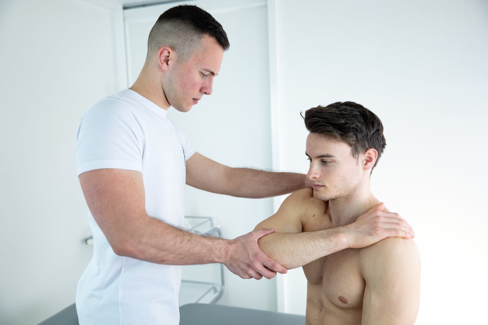
Shoulder joint is built so that the cup looks like a shallow saucer, and is located on the scapula and is called the glenoid, and the joint head is an almost regular sphere, significantly larger than the cup. Such a structure allows great mobility, but also considerable instability.