
Imaging diagnostics, especially MRI, has brought significant benefits to orthopedics and physiotherapy. Especially today, when examinations with 3T resolution machines are available to most. Whenever we are unsure about a diagnosis, MRI is there, and the information it provides is usually useful. However, relying predominantly on images and less on physical examination has its drawbacks.
The advantages of ultrasound over magnetic resonance imaging
Description
Patients themselves prefer having “pictures” showing their problem. Unfortunately, MRI is not entirely accurate. One of our most eminent radiologists says that MRI is accurate in about 80% of cases, and that’s if it’s interpreted by a very experienced specialist. Besides this fact, we can compare MRI to a regular photograph. It is static, showing only one moment in your life and in only one position. Even a photograph taken 2 hours later will be different, and in a different position, additional differences will be seen. Also, a photograph shows the shape or form but cannot say anything about function. So based on your photograph, no matter how good the resolution is, I cannot conclude whether you can run and for how long, whether you can jump, or whether you can walk. The same applies to MRI scans. Without a proper examination, their significance is not significant.
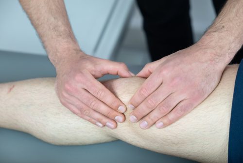
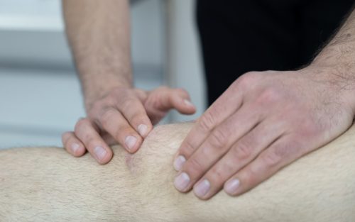
A recent example vividly illustrates this. A female patient sought our help due to persistent pain on the outer side of her left knee. MRI showed minor damage to the lateral meniscus, and the orthopedist recommended arthroscopy, which she underwent shortly thereafter, during which the damaged part of the meniscus was removed. However, after a short recovery from the operation, her pain remained unchanged. That’s when she came to us. It is significant that about 15 years ago, she fell on that knee, limped for a while due to it, and hit precisely the spot that hurts her today. A simple ultrasound examination revealed a small cluster of calcifications in the area of the thigh bone, resulting from that fall, and the tissue around them became inflamed, causing pain. Several simple therapies later, and she is now completely pain-free. Unfortunately, MRI often does not detect such calcifications.