
Literally the entire medical profession and all patients, together with us, view knee osteoarthritis as damage to or wear of the cartilage in the joint. Although this observation is correct, the question remains what we can do regarding such damage, and the answer is very simple—absolutely nothing.
Knee osteoarthritis
Description
In the final stage of the disease, it is possible to surgically implant an artificial joint, and that is all. All injections that promise “regeneration” of cartilage only reduce pain, which is good and desirable, but they do not repair damage to the cartilaginous surface, and arthroscopic joint debridement serves the same purpose. All physiotherapy is of a general type and is directed toward reducing inflammation and “strengthening” muscles, without a clear understanding of what led to their weakness. During the course of the disease, it is common for knee pain and swelling in the first two stages of osteoarthritis to appear and disappear in waves, independently of the patient’s activities and environment, which we cannot fully explain; in short, we know much about osteoarthritis, but we assume even more.
In this context, our proposal is unusually simple. For a moment, let us put cartilage aside and view the osteoarthritic knee as a whole, where the first thing that becomes evident is joint instability similar to that seen in ligament injury or rupture. In such instability, swelling and pain also occur intermittently and disappear spontaneously, and if it persists long enough, it leads to cartilage damage similar to that seen in osteoarthritis. The only difference is that in knee instability caused by ligament damage, we have a clear understanding of what can and should be done to make the knee more stable, reduce swelling and pain, and protect cartilage from further damage.
This leads to a simple question: can we observe knee osteoarthritis through the prism of instability and in that way make physiotherapy logical, unidirectional, and effective, and if we can, why do we not do so? For all those who wish to better understand their own or another person’s problem and effectively help themselves or others without sensationalist phrases characteristic of modern communication on social networks, we offer some answers and numerous explanations, and therefore we proceed step by step.
All forms of osteoarthritis are similar in their physiology and pathoanatomical changes, and more information about them can be found on our website. Nevertheless, osteoarthritis of each joint is different because their function, load level, and role in movement differ, which also implies differences in treatment, particularly in physiotherapy and kinesiotherapy. This is also the case with the knee.
Joints can be classified in various ways, but for this discussion we divide them into three groups: joints that bear large loads and express strength, such as the hip, spine, and shoulder; joints that primarily serve a shock-absorbing function, such as the knee and ankle; and joints responsible for precise movements, such as the hand and elbow. None of these joints has only one function, but one function can be considered dominant in the complex task of the locomotor system. Consequently, osteoarthritis of each of these joints affects function differently, and the methods for maintaining, restoring, or preventing degradation must differ; in short, although the anatomical process of osteoarthritis is similar in all joints, treatment methods, especially physiotherapy, must necessarily differ if optimal therapeutic effects are to be achieved.
Knee osteoarthritis has four stages—initial, mild, moderate, and terminal—between which the differences are subtle and without a clearly defined transition. These stages are determined by a combination of symptoms, such as pain, swelling, and restricted mobility, and imaging findings obtained through X-ray and magnetic resonance imaging, and they primarily serve to guide the selection of therapeutic interventions aimed at reducing pain and improving mobility. Another important fact is that knee osteoarthritis has spontaneous phases of worsening and improvement without an apparent external trigger, which occur due to internal processes within and around the joint through which the body attempts compensation via biological and biomechanical changes, including changes in bone shape (osteophytes), as well as changes in cartilage, the joint capsule, and ligaments.
These phases may coincide with certain therapeutic interventions, creating the impression of their effectiveness, while in reality they may have been applied during a period of spontaneous improvement, which forms the basis of many urban legends about miraculous teas, creams, ointments, herbal preparations, and other alternative treatments. While there is nothing inherently wrong with alternative or complementary medicine, this explains why such methods do not always work or lose their effect over time.
Conventional physiotherapy generally proceeds in two directions: one aimed at reducing inflammation and swelling through instrumental physiotherapy, including electrical stimulation, ultrasound, magnetic therapy, radiofrequency therapy, pressotherapy, and, in specific cases, shockwave therapy, and the other aimed at strengthening thigh muscles weakened by pain and swelling through exercises designed to activate muscles while avoiding excessive loading of damaged cartilage, supplemented by proprioceptive exercises intended to improve muscle activation timing and thereby enhance joint protection during movement. The underlying philosophy of this approach is the treatment of visible symptoms and their consequences in the hope of improving quality of life, which indeed occurs in some patients, most commonly in the early stages of the disease, whereas in later stages this approach becomes less effective and requires a different conceptual framework.
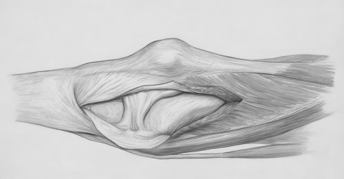
The tension model
The knee is predominantly a shock-absorbing system of considerable complexity and can be simplified as a spring designed to reduce stress during walking, running, jumping, and changes of direction by absorbing forces that would otherwise be transmitted to the rest of the body. This spring functions properly only when all its components are intact and in proper mutual tension, or tensional equilibrium, and when this equilibrium is disrupted, shock absorption is reduced and eventually collapses, as is the case in osteoarthritis. Such a collapse can be visualized as a partially damaged spring that becomes unstable, with every applied force causing further instability and damage.
While replacing the spring is not possible in the case of the knee, it is possible to place the damaged spring into a stabilizing housing that reduces lateral instability, thereby improving shock absorption and slowing further damage, which is precisely the goal in treating the osteoarthritic knee. Instability in the knee can be static or dynamic, with dynamic stability provided by muscles and their ability to activate at the correct time, and static stability provided by ligaments, which are connective tissues designed to limit excessive joint movement. Ligament tension depends on cartilage thickness, and as cartilage thins in osteoarthritis, ligaments become looser, reducing static stability and leading to micro- and macro-instability, which further damages cartilage and perpetuates a vicious circle.
A solution does exist, as muscles can form a new stabilizing structure around the knee, but understanding this requires insight into the evolutionary mechanism that leads to muscle weakening in unstable joints. Following knee injury, the body limits muscle strength through a mechanism known as the small reflex arc, which evolved to protect acutely injured tissue but becomes maladaptive in chronic conditions such as osteoarthritis, where it remains permanently active and progressively weakens surrounding muscles. Attempts to increase absolute muscle strength without addressing this mechanism often exacerbate instability, pain, and swelling, which explains why exercise may worsen symptoms in some patients.
Understanding that muscle weakness is a consequence rather than a cause of joint damage allows for the development of exercise programs that bypass rather than stimulate the small reflex arc, using physiotherapy techniques to modulate sensory input and specially designed exercises to reduce pain and increase muscle strength without reactive symptom exacerbation. This approach improves both static and dynamic knee stability, slows further cartilage degeneration, and improves quality of life, although its successful application requires time, knowledge, and patient consistency, which remains its primary limitation in the context of widespread promises of effortless and rapid cures.
RELATED CONTENTS

Premium
Plaćeno
The main characteristic of this condition is pain in the knee area, specifically just below or above the patella (kneecap). This is the small bone located at the front of the knee, easily noticeable as it lies directly beneath the skin. Its function becomes clearer when understanding its position within the knee joint.

Premium
Plaćeno
Imaging diagnostics, especially MRI, has brought significant benefits to orthopedics and physiotherapy. Especially today, when examinations with 3T resolution machines are available to most. Whenever we are unsure about a diagnosis, MRI is there, and the information it provides is usually useful. However, relying predominantly on images and less on physical examination has its drawbacks.

Premium
Plaćeno
Anterior knee pain is common in adolescents and young women, often with no clear diagnosis. Discover possible causes and treatment approaches using the holistic tension model.