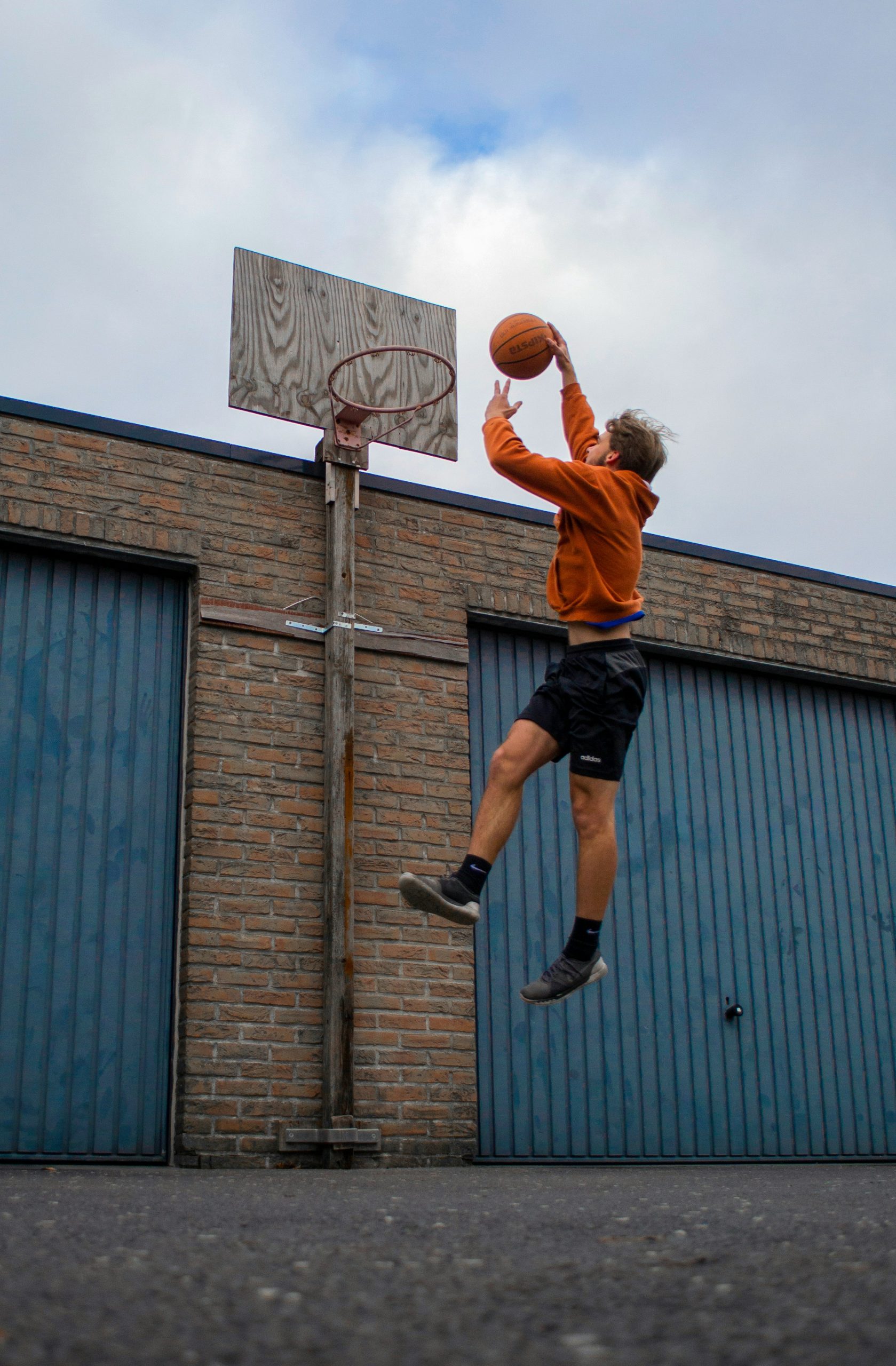
True jumper’s knee is characterized by pain on movement initiation and tenderness located below or above the patella. This pain occurs during physical exertion and after it, and it is marked by long duration and weak responsiveness to existing therapeutic methods. However, there are situations in which typical soreness is present, and ultrasound shows tendon changes in the typical location, with one exception — a small inflammation visible as weak or absent Doppler activity. An additional difference is that rest from physical activities rarely brings relief, and the location of pain can change, although within a relatively small spatial area. In a portion of false jumper’s knees, pain can vary in intensity, regardless of physical activity, or completely withdraw for shorter periods of time, only to reappear in full strength, also regardless of physical exertion. One of the most important diagnostic procedures for distinguishing true from false jumper’s knee is also a therapeutic procedure, namely mobilization, that is, manipulation of the patellofemoral joint — the connection between the patella itself and the groove on the femur along which the patella slides during movement and physical and sports activities. If it is a false jumper’s knee and if the procedure on the patella is performed precisely, instant symptom reduction occurs, sometimes even their complete elimination. This is not possible with true jumper’s knee.
If you try to search scientific papers or literature that discusses this syndrome, you will not be very successful. Everything described so far is exclusively the result of our many years of experience and exchange of information with colleagues from the Netherlands, Poland, Portugal and the United Kingdom and as such serves for your information, but does not represent a scientific fact. At least not yet.
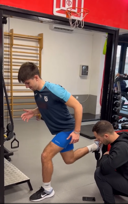
This condition and the possible causes of pain similar to patellar apicitis can be viewed in three ways. One is a change in the joint itself between the patella and the femur, which may include softening or cartilage damage, or dysplasia, that is, a poor shape of joint bodies that allows micro or macro instabilities during movement and sports, so the local pain at the tip of the patella is only transferred from the underlying disease. The second are changes in the soft tissues and ligaments that surround the knee and provide stability to it, but change with the type of physical activity or lack thereof, and the local and global tensions of those same tissues are altered, projecting pain to the tip of the patella in that way. The third are distant causes, such as, for example, hip stiffness, previously repeated ankle distortions with resulting local instability, or chronic low back pain. Each of these, as well as many other unmentioned conditions, affect the role of the extensor knee system, of which the patella is an integral part, in such a way that they place demands on it for which it did not evolve, and in the specific case it is not sufficiently conditioned. The manipulation of the patella itself, which we use for diagnostic and therapeutic purposes, results in an instant tension change of the structures of the extensor knee system, and in that way reduces or eliminates pain during movement. Such a result tells us that the altered tension of connective structures is precisely the direct cause of pain, and not the existence of a true pathological process. Just like with any stretching, if it lasts long enough, the consequence will be pain, which usually decreases or stops when the stretching stops. With the cessation of increased tension, the pain also stops.
Although manual therapy, in this case on the patellofemoral joint, can be and is a powerful lever in the treatment of many movement system disorders, it is rarely sufficient for the complete rehabilitation of false jumper’s knee.
The tension model is already at the very basis for the distinction of this painful syndrome as a separate entity. It predicts pain localization distance from its true cause, through a change in tension in the tissues of the entire extremity or the entire movement system. In that sense, it provides tools for precise diagnostics of false jumper’s knee, as well as for determining local or distant causes of its occurrence. When this is done, treatment is generally fast, efficient, individually modeled, with clear instructions for its further prevention, and in addition to manual therapy includes an individualized exercise program with the aim of remedying identified deficiencies.