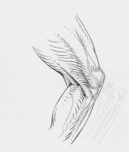
The main characteristic of this condition is pain in the knee area, specifically just below or above the patella (kneecap). This is the small bone located at the front of the knee, easily noticeable as it lies directly beneath the skin. Its function becomes clearer when understanding its position within the knee joint.
Jumper’s Knee
Description
It is “inserted” into the tendon of the large thigh muscle (quadriceps or four-headed thigh muscle) and serves as a movable fulcrum point, thus reducing tendon friction during movement and facilitating the movement itself. During heavy loads over a prolonged period, cumulative damage and subsequent inflammatory processes can occur precisely at the points where the tendon attaches to the patella or shinbone, and then this condition is called “jumper’s knee.”
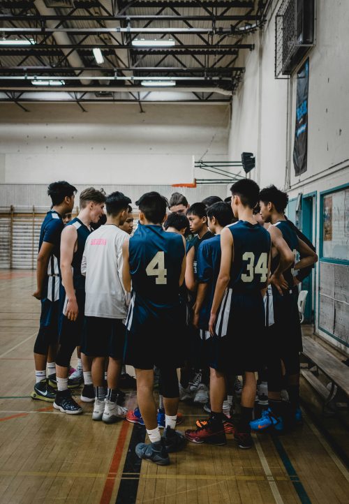
This painful syndrome is a common occurrence in sports that involve frequent jumping, running, or knee extension with significant loads. For example, in volleyball, jumper’s knee accounts for 28% of all injuries, with as much as 40% of elite volleyball players experiencing problems during their sports careers. The percentages are only slightly lower in basketball, handball, athletics (high jump, long jump, triple jump, sprint), weightlifting, cycling, figure skating, ice hockey, skiing, cross-country skiing, or tennis. All this puts this overuse syndrome in an inglorious first place in terms of frequency, which does not apply only to professional sports but also to recreational athletes.
The location of pain, and thus inflammation, does not necessarily have to be at the so-called apex of the patella. There are three locations where jumper’s knee can manifest. One is at the already mentioned tip of the patella, accounting for about 75%, the second is above the patella, or at its base, accounting for about 18%, while the third is at the attachment of the patellar ligament (the extended part of the quadriceps tendon) to the shinbone, accounting for about 7% of all cases of jumper’s knee. These figures vary from study to study.
 In addition to pain on pressure and during squatting, typical is the pain that occurs during warm-up, partially subsides or completely disappears during training, only to reappear and intensify at the end or after training or a match. The first steps after getting out of bed following a night’s rest can be painful, as can walking up and down stairs. Rest from training for a few days may completely eliminate the pain, only for it to reappear with the first subsequent physical strain. When such symptoms appear, it is advisable to perform additional examinations to objectify the findings, and this is best done with ultrasound diagnostics, because, in addition to confirming the clinical diagnosis, it is important to determine the stage of the disease. Since the pathological process is located just under the skin, modern ultrasound machines produce images comparable to MRI, are significantly cheaper than other imaging diagnostics, and provide real-time information. Additionally, ultrasound allows for dynamic examination during muscle tension or knee movement, which is not possible with other methods. All this provides important information about:
The extent of the damage
The location of the damage
The size and form of the inflammatory process
Armed with the results of the clinical examination and ultrasound diagnostics, we are able to predict the approximate duration of treatment, the type of therapeutic procedures needed, the likelihood of treatment success, and the need for additional interventions in the field of orthopedics.
Like any other overuse syndrome, jumper’s knee is not an inflammation in its basic sense, which includes four stages: acute inflammation, subacute state, primary healing, and remodeling, ending with complete repair of the damage. Instead, in the chronic state, this inflammatory cascade “gets stuck” halfway and cannot complete healing, which is why symptoms and tissue condition do not improve even after a long time. It is as if the entire process has entered a dead-end and cannot proceed further.
The usual approach to treatment involves the application of a full range of physiotherapy procedures, from laser, electrotherapy, radiofrequency therapy, magnetotherapy, manual therapy methods, with an emphasis on mobilization of the tendon and surrounding soft tissues, combined with exercises aimed in three directions: strengthening weakened muscles, stimulating reflex response speed, and stretching.
If such an approach is insufficient, we introduce shockwave therapy, which provokes damaged tissue with sound bursts and induces a new, acute inflammation, with the aim of initiating a new inflammatory cascade reaction that should result in final healing. This is an extremely powerful and effective method when applied to appropriate cases and in combination with other therapeutic procedures.
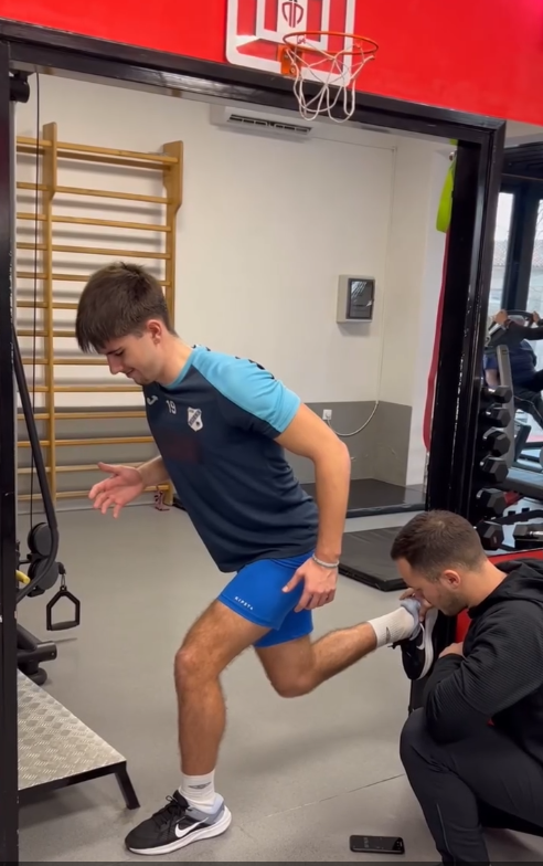
Additionally, in recent years, an attempt has been made to apply isolated eccentric contraction (link to article), which has proven to be an excellent intervention in chronic Achilles tendon inflammation. The idea is to stimulate changes in damaged tissue with only one type of movement, in the full range of motion, and with maximum load. The problem with its application to the knee is the patella and its cartilage, which are under extremely high pressure and stress during isolated eccentric contraction. Since its damage underlies most jumper’s knee cases, this often means that by treating one condition, we worsen another. Therefore, this method can be an option and addition in treating jumper’s knee but not in all cases, and then under professional guidance and with careful application aimed at minimizing its negative consequences.
Simplified, there are two therapeutic approaches.
The first aims at reducing inflammation and pain, with exercises to activate targeted muscles, while the second is stimulatory and aims at provoking new inflammation, along with addressing specific deficiencies in surrounding soft structures. Both aim to conclude the chronic inflammatory process with healing and prepare the tendons and knee for full load.
As can be seen from available literature, neither approach is successful in more than 60-70% of cases, even when performed over a sufficiently long period. The reason for their relative success rate is always the same—they view the knee and quadriceps tendon as isolated parts of the body, almost without any connection to other integral parts of the locomotor system. Although jumper’s knee can be a problem only of the knee and its dysfunction, in reality, this is rarely the case. Furthermore, both approaches exclude individual differences between patients, which not only can but actually do play a crucial role in the development of every overuse syndrome, including jumper’s knee.
The tension model (link to article) fundamentally postulates the individual variability surrounding every diagnosis. A simple example is a blister on the foot that appeared after prolonged walking or running. The blister itself, by definition, is an overuse syndrome of the skin due to intense friction. But whether that blister arose due to too tight or too loose shoes, poor socks, moist skin, the specific shape of the heel bone, prolonged walking or running, the technique of walking or running, uneven terrain, poorly tied shoelaces, some other reason, or a combination thereof is the fundamental question of every clinical process that includes not only an examination of the local blister, in this example, but also an examination of the entire locomotor system, possible imaging diagnostics, medical history, and a deep insight into the habitual component of the overuse syndrome’s occurrence, i.e., the patient’s habits themselves. Only when all the pieces of the puzzle are in place is it possible to have a clear picture of the painful condition’s origin in a specific case, and then the intervention is logical, individually modified, and aimed at addressing the specific deficiencies observed. This picture is unique to that patient and no one else. The protocols we use today are generalized standardizations that treat the patient as one and the same type of car, where one and the same malfunction is repaired in one and the same way. In the specific case of jumper’s knee, local pain and inflammation are similar to a blister on the foot. It is the same disease or painful condition for all who suffer from it, but the reason for its occurrence is fundamentally different for each of them. Without the tools of the tension model or a similar system of individualized diagnostics and therapy, we are left at the mercy—and more often the lack of mercy—of protocols and are partially or completely powerless when one or more protocols prove unsuccessful in an individual case.
Additional information:
Cortisone injections (a synthetic hormone with strong anti-inflammatory effects) are avoided in jumper’s knee as they significantly increase the risk of tendon rupture.
Hyaluronic acid injections, recently used, have not yet shown significant benefits in treating this overuse syndrome.
PRP injections (isolated blood plasma) can, according to some studies, partially shorten the duration of treatment in some patients but do not diminish the importance of the diagnostic and therapeutic process described in this text.
Surgical treatment is now very rare and makes sense only when the pathological change is so large and so resistant to physiotherapy that it remains the only solution. It involves removing the damaged tendon attachment along with part of the bone it attaches to. It is successful in a high percentage of cases, but postoperative recovery, especially until readiness for full training and sports load, is very long.


